
TLDR
- The most common examples of X-rays are chest films (lungs, heart), bone films (fractures, arthritis, spine), dental films (cavities, wisdom teeth), mammograms (breast screening) and contrast studies such as the barium swallow.
- Outside medicine, X-rays check airport luggage, inspect shipping containers, expose hidden layers in paintings, reveal crystal and DNA structure, and map black holes from orbit.
- A single chest X-ray delivers roughly the radiation you’d pick up from background sources in two to twelve days. A CT scan of the same area delivers about 70 times more.
Table of contents
- How X-rays make a picture
- Medical examples of X-rays
- Dental examples of X-rays
- Beyond medicine: X-rays in security, art and space
- X-ray vs. CT vs. fluoroscopy
- Radiation dose: how much is a typical X-ray?
- What to expect at an X-ray appointment
- Are X-rays safe?
An X-ray is a shadow picture. A machine sends a brief burst of high-energy light through your body, and a detector on the other side records how much made it through. Dense material like bone stops most of it and shows up white. Air in the lungs stops almost none and shows up black. Everything else lands somewhere in between.
That one principle produces a surprising range of images. Here are ten examples of X-rays you’re most likely to meet in a clinic or dentist’s office, followed by the less obvious ones: the scanner at airport security, the machine that found a hidden portrait under a Van Gogh, and the telescope that photographs black holes.
How X-rays make a picture
Wilhelm Röntgen produced the first X-ray image in 1895, a photograph of his wife’s hand with her wedding ring visible as a dark band around the finger bones. He won the first Nobel Prize in Physics for it in 1901. The basic physics hasn’t changed since.
X-rays are electromagnetic radiation, the same family as visible light and radio waves, but with wavelengths short enough to pass through soft tissue. The more calcium, metal or other dense material in the way, the more X-rays get absorbed, and the whiter that region looks on the image. This is why X-rays are the standard first test for suspected broken bones: bone is the densest natural thing in your body.
Medical examples of X-rays
Medical imaging accounts for most of the X-rays taken worldwide. These are the first eight, ending with a screening test and a contrast study.
1. Chest X-ray
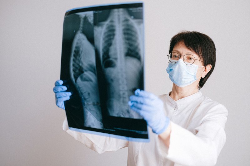
The most common X-ray in the world. You stand facing a flat detector, take a breath in, and hold it for a second. The radiologist reads the lungs, the outline of the heart, the diaphragm, and the ribs in one image.
Healthy lungs look dark because they’re full of air. Pneumonia shows up as a white patch where fluid has replaced that air. A collapsed lung appears as a dark rim of air outside the lung edge, and an enlarged heart shows as a silhouette that fills more than about half the width of the chest. Doctors order it for a persistent cough, chest pain, shortness of breath, and before many surgeries.
2. Fracture (bone) X-ray
The textbook example. A broken bone appears as a dark line cutting through a white one, or as a step where the bone’s edge no longer lines up. Technicians usually shoot two or three angles, because a crack running directly toward the detector can vanish in a single view.
One quirk: tiny stress fractures, common in runners’ feet and shins, often don’t show up on the first film. If the pain persists, a repeat X-ray a week or two later can reveal the callus, the new bone the body lays down to repair the crack.
3. Joint X-ray for arthritis
X-rays can’t see cartilage directly, but they can see the gap it leaves. In a healthy knee, the space between the thigh bone and shin bone is even and clear. In osteoarthritis, that gap narrows as cartilage wears away, and small bony spurs called osteophytes grow along the edges. Knee and hip films are often taken standing, since body weight squeezes the joint and exposes narrowing that a lying-down image would hide.
4. Spine X-ray for scoliosis
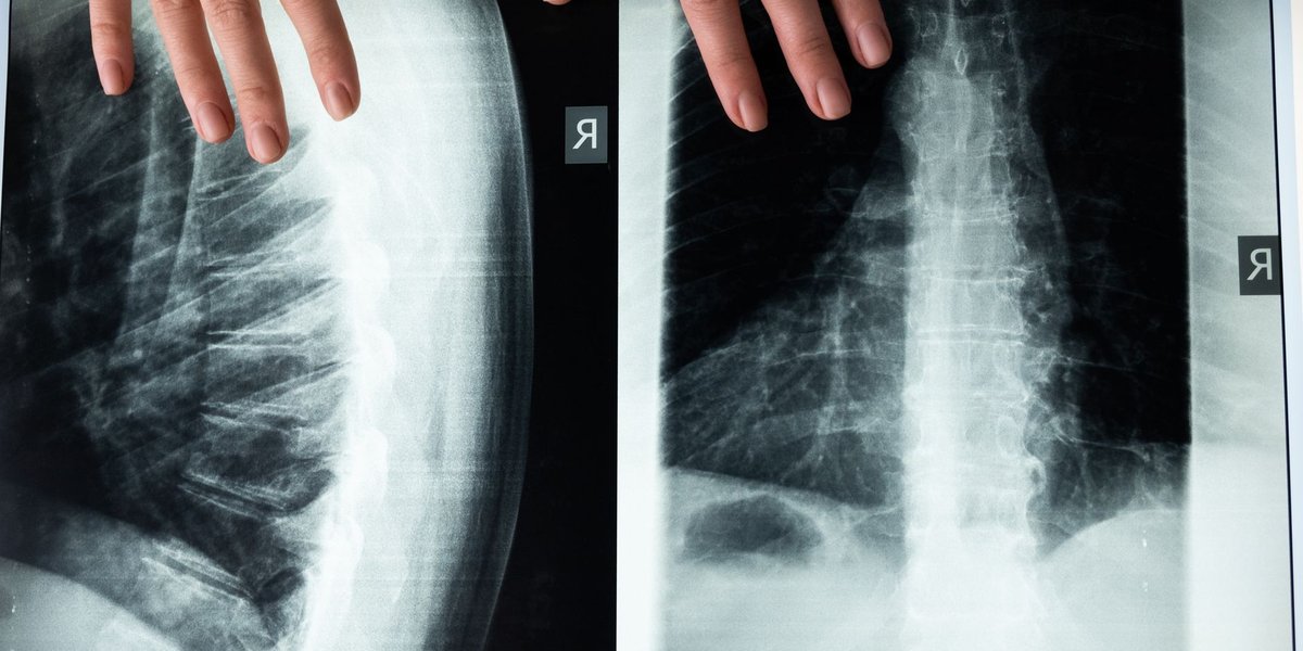
A full-length image of the spine lets the radiologist measure its curve. The standard measure is the Cobb angle: lines are drawn along the tilted top and bottom vertebrae of the curve, and the angle between them is the score. A curve of 10 degrees or more counts as scoliosis. Children with a growing curve get repeat films every few months so doctors can see whether it’s progressing.
5. Hand and wrist X-ray for bone age
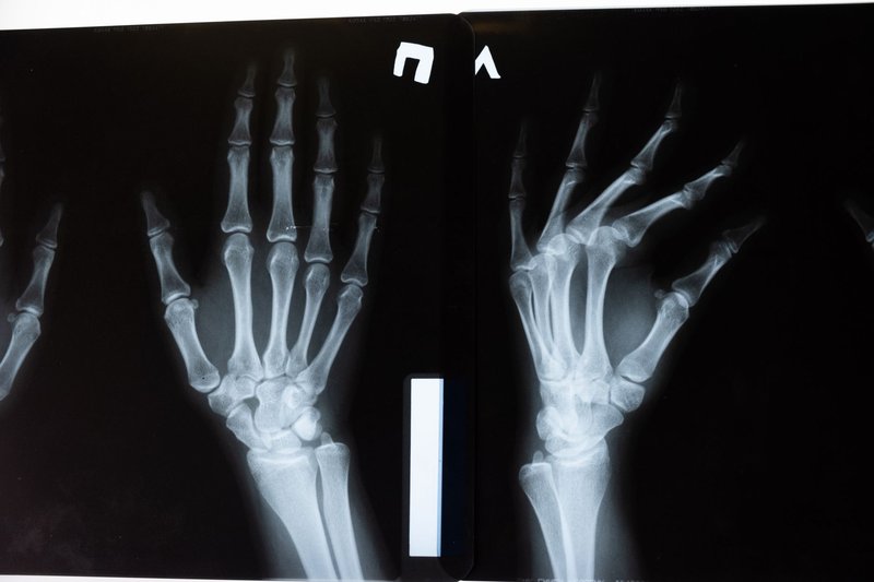
This one surprises people. Growth plates, the soft zones at the ends of a child’s bones, close at predictable ages. A film of the left hand and wrist shows how far along that process is, so a doctor can compare a child’s skeletal age with their calendar age. A 10-year-old with the hand bones of a 7-year-old, or a 13-year-old, points toward a growth or hormone problem worth investigating.
6. Abdominal X-ray
Often called a KUB (kidneys, ureters, bladder). It’s a quick film for suspected bowel obstruction, where loops of gas-filled intestine stack up in a telltale pattern, and for kidney stones that contain enough calcium to show as small white specks. It’s also the go-to image when a toddler swallows a coin or a button battery, since metal is easy to spot.
7. Barium swallow (contrast X-ray)
Soft organs like the esophagus are nearly invisible on a plain film, so radiologists add contrast. In a barium swallow, you drink a chalky barium sulfate liquid. Barium absorbs X-rays strongly, so as it coats the throat and esophagus, the outline glows white against the darker background. The test shows narrowing, pouches, or reflux, and the radiologist usually watches in real time as you swallow. Merck Manuals describes the same approach for the stomach and intestines, where the barium is swallowed or given as an enema.

8. Mammogram
A mammography machine compresses the breast between two plates and uses a lower-energy X-ray beam, tuned to separate subtle differences between fatty and dense tissue. The compression is uncomfortable for a few seconds, but it spreads tissue out so small abnormalities, including tiny calcium deposits that can signal early cancer, don’t hide behind each other. In the US, the Preventive Services Task Force recommends screening every two years for women from 40 to 74.
Dental examples of X-rays
Dentists were early adopters, and a typical dental X-ray uses a tiny fraction of the dose of a body film.
9. Bitewing X-ray
You bite down on a small plastic tab holding the sensor, and the image captures the crowns of your upper and lower back teeth in one frame. It’s built for one job: finding cavities between teeth, where no mirror or probe can reach. Early decay looks like a small dark wedge in the white enamel. Bitewings also show the level of the bone supporting your teeth, which drops with gum disease.
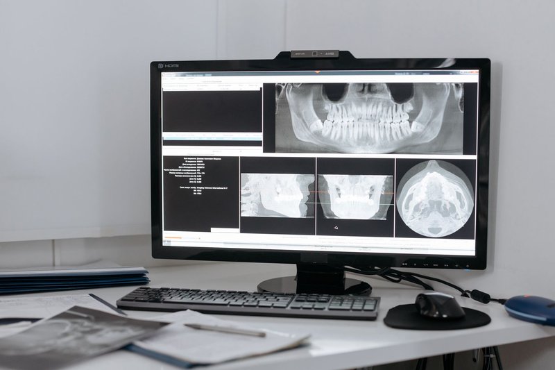
10. Panoramic X-ray
A machine rotates around your head over about 20 seconds and produces a single wide image of both jaws, all your teeth, the sinuses and the jaw joints. This is the film dentists and oral surgeons use to plan wisdom tooth removal, because it shows how the roots sit relative to the nerve canal in the lower jaw. It also helps with orthodontics and implants.
Beyond medicine: X-rays in security, art and space
The same shadow principle works on anything dense enough to absorb X-rays.
Airport baggage scanners. Checked and carry-on bags pass through a machine that images them from one or more angles. The software colors the image by material: orange for organic items like food and clothing, blue for metals, and green for mixtures. Operators learn to recognize the dense, wire-laced shape of a device that doesn’t belong. The body scanner you walk into is a different technology. US airports use millimeter-wave scanners, after retiring X-ray backscatter machines in 2013.

Cargo and truck inspection. Border agencies run shipping containers and entire trucks through gantry-sized scanners using high-energy X-rays or gamma rays. A container full of identical boxes has an even, repeating pattern. A hidden compartment or a stack of something denser disrupts it.
Art authentication and restoration. Lead white, a pigment painters used for centuries, absorbs X-rays strongly. An X-ray of a painting therefore reveals the structure of the lead-based layers underneath the surface, including earlier compositions the artist painted over. A portrait of a woman hidden beneath Van Gogh’s Patch of Grass was found this way. Conservators also use X-rays to check for repairs and to flag forgeries painted on old canvases with modern materials.
Crystallography. When X-rays pass through a crystal, they scatter into a pattern of spots that encodes how the atoms are arranged. Max von Laue demonstrated this in 1912, and in 1952 Rosalind Franklin’s X-ray diffraction image of DNA, known as Photo 51, gave Watson and Crick the evidence for the double helix. Today the same technique maps the structure of proteins and drug targets.
X-ray astronomy. Earth’s atmosphere blocks X-rays from space, which is good for us and bad for astronomers, so telescopes have to fly. NASA’s Chandra X-ray Observatory, launched in 1999, images gas heated to millions of degrees around black holes, colliding galaxy clusters, and exploded stars. The first X-ray source found beyond our solar system, Scorpius X-1, was discovered in 1962 by a rocket-borne detector.
X-ray vs. CT vs. fluoroscopy
These get lumped together, but they’re different tools, and the difference matters when you’re comparing a chest X-ray vs. a CT scan.
| Plain X-ray | Fluoroscopy | CT scan | |
|---|---|---|---|
| What it produces | One flat still image | Live X-ray video | Cross-section “slices,” combined into 3D |
| Typical use | Fractures, pneumonia, dental | Barium swallow, guiding catheters | Internal bleeding, tumors, complex fractures |
| Relative dose | Low | Moderate to high | High |
A CT scanner rotates an X-ray source around you and rebuilds hundreds of angles into thin slices, so a small nodule that a chest film would miss behind a rib shows up clearly. The price is dose. Fluoroscopy is the opposite trade: a continuous beam for seconds or minutes, which is how a radiologist watches barium move down your esophagus or guides a wire through a blood vessel.
If a doctor offers a plain X-ray first, it’s usually because it answers the question for a fraction of the radiation and cost. CT is the follow-up when the film leaves something unresolved.
Radiation dose: how much is a typical X-ray?
Dose is measured in millisieverts (mSv). The average person in the US absorbs about 3 mSv a year from natural background sources such as radon in soil, cosmic rays and trace radioactivity in food, according to the EPA. That works out to roughly 8 microsieverts a day, which gives an easy yardstick: how many days of ordinary life equals this scan?
| Exam | Typical dose (mSv) | Roughly equal to |
|---|---|---|
| Hand or foot X-ray | 0.001 | About 3 hours of background |
| Dental bitewing set | 0.005 | About 14 hours |
| Chest X-ray | 0.02 to 0.1 | 2 to 12 days |
| Mammogram | 0.4 | About 7 weeks |
| Abdominal X-ray | 0.7 | About 3 months |
| Upper GI series (barium) | 6 | About 2 years |
| Chest CT scan | 7 | About 2 years |
The chest X-ray range is wide because a modern digital single-view film sits at the low end and an older or two-view exam sits higher. Doses vary by machine, body size and technique, so treat these as ballpark figures.
What to expect at an X-ray appointment
The visit is short. For a plain X-ray, expect about 10 to 20 minutes door to door and a few seconds of actual exposure.
- Clothing and jewelry. You’ll change into a gown or remove clothing and jewelry over the area, since metal blocks the beam and leaves shadows on the image.
- Positioning. The technician arranges you against the detector and steps behind a shield. You’ll be asked to hold still, often to hold your breath.
- Pregnancy. Tell the technician if you might be pregnant. Shielding or a different test may be used.
- Contrast studies. For a barium test, you may need to avoid eating for several hours beforehand, and the chalky drink is the least pleasant part. Expect pale stools for a day or two afterward.
- Results. A radiologist reads the images and sends a report to your doctor, typically within a day, faster for emergencies.
Are X-rays safe?
For a single diagnostic X-ray, yes. The radiation is ionizing, meaning it can damage DNA in principle, but the doses from plain films are comparable to a few days of natural background exposure, and the diagnostic benefit of finding a fracture, infection or tumor early outweighs the very small added risk. The risk adds up with repeated high-dose scans such as multiple CTs, which is why doctors follow a rule of using the lowest dose that answers the clinical question and skipping imaging that won’t change the treatment plan.
If you’re asked to get an X-ray and wonder whether it’s necessary, ask what decision the result will change. That question is reasonable, and any good clinician will have an answer.