

TLDR
Flat bones are thin, curved, plate-like bones built for protection and muscle attachment rather than leverage. The main examples: the frontal, parietal, occipital, nasal, lacrimal, and vomer bones of the skull; the sternum and ribs of the thoracic cage; the scapula; and the ilium, ischium, and pubis that fuse into each hip bone. Depending on how you count ribs and hip bones, that’s somewhere around 36 to 38 flat bones total — more on that math below.
Table of Contents
- What Makes a Bone “Flat”
- Flat Bones of the Skull
- Flat Bones of the Thoracic Cage
- The Scapula
- Flat Bones of the Pelvis
- How Flat Bones Are Built
- What Flat Bones Actually Do
- How Many Flat Bones Are in the Body
- Flat Bones vs. the Other Three Types
- Where This Matters Clinically
- Quick Self-Check
What Makes a Bone “Flat”
A flat bone is thin, often curved, and shaped more like a shield than a rod. That’s the whole design philosophy: long bones (femur, humerus) are built to act as levers for movement, and flat bones are built to cover something or give muscle a wide surface to grab onto. No shaft, no rounded ends, no joint cavity doing the work — just a broad plate of bone doing a plate’s job.
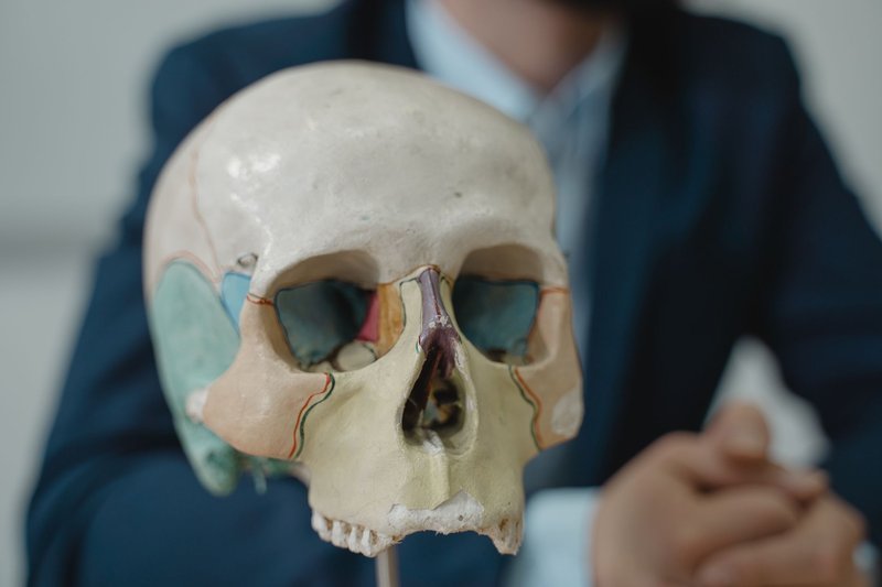
Anatomists sort every bone in the body into five shape categories: long, short, flat, irregular, and sesamoid. Flat bones make up a smaller slice of the skeleton than long bones do, but they cover the parts of you that really can’t afford a structural failure — your brain, your heart, your pelvic organs.
Flat Bones of the Skull
| Bone | Count | What it protects or anchors |
|---|---|---|
| Frontal | 1 | Forehead and upper eye sockets; shields the frontal lobes |
| Parietal | 2 | Top and sides of the skull, forming the crown |
| Occipital | 1 | Back and base of the skull; the foramen magnum runs through it for the spinal cord |
| Nasal | 2 | Bridge of the nose; anchors cartilage that shapes the rest of it |
| Lacrimal | 2 | Smallest bones in the face, sitting at the inner corner of each eye socket; house part of the tear drainage system |
| Vomer | 1 | Thin blade of bone forming the lower back of the nasal septum |
That’s nine flat bones in the skull alone, and they’re a genuinely elegant piece of engineering. At birth, several of them — the two frontal, two parietal, and one occipital — aren’t fused yet. They’re separated by soft connective-tissue gaps called fontanelles, the “soft spots” every parent gets warned about. According to MedlinePlus, the posterior fontanelle typically closes within the first two to three months of life, while the anterior one — the largest — can stay open anywhere from seven months to nearly two years. That flexibility isn’t a design flaw; it’s what lets an infant’s skull compress slightly during birth and then keep expanding as the brain triples in size over the first year.
Flat Bones of the Thoracic Cage
The chest gets two kinds of flat bone, and together they form a cage rather than a shield:
- Sternum — the breastbone, a single flat plate running down the center of the chest where the ribs anchor via cartilage
- Ribs — 24 total (12 pairs), each one a long, thin, curved flat bone rather than the rod-shaped long bone people sometimes assume it is
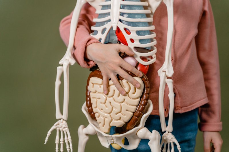
Ribs get miscategorized a lot because “rib” sounds like it should be a long bone — it’s long and curved, after all. But structurally a rib is built the same way a skull plate is: two layers of dense compact bone sandwiched around a spongy core, not a shaft with a marrow cavity running down the middle. That’s the test that actually matters for classification, not the overall silhouette.
The Scapula
The shoulder blade is one of the few flat bones that sits almost entirely on its own, out on a limb rather than boxed into a cage. It’s a broad, triangular plate on the back of the ribcage that gives more than a dozen muscles — trapezius, deltoid, rhomboids, rotator cuff muscles — somewhere to attach. You have two, one per side, and they float relatively freely, tethered mostly by muscle rather than rigid joints, which is exactly why your shoulder has the largest range of motion of any joint in the body.
Flat Bones of the Pelvis
Each hip bone (the correct name is the “os coxae,” though most people just say “pelvis”) starts life as three separate flat bones that fuse together by early adulthood:
| Bone | Region | Role |
|---|---|---|
| Ilium | Upper, wing-shaped portion | Forms the hip you feel when you put your hands on your waist; anchors major glute and abdominal muscles |
| Ischium | Lower-back portion | Bears your weight when you sit — the “sit bones” |
| Pubis | Lower-front portion | Meets its counterpart at the pubic symphysis, closing the pelvic ring in front |
By your mid-20s, the cartilage between these three has ossified completely, and what was three bones reads as one on an X-ray. Anatomists still count them as three distinct flat bones for classification purposes, because that’s how they form and how they’re described in every anatomy course.
How Flat Bones Are Built
Cut a cross-section through any flat bone and you’ll find the same three-layer sandwich every time:
- Periosteum — a thin, fibrous outer membrane wrapping the whole bone, packed with nerve endings and blood vessels and serving as the attachment point for tendons
- Compact bone — a dense outer and inner layer, hard and smooth, providing the structural strength
- Spongy (cancellous) bone — a porous, honeycomb-like core sandwiched between the two compact layers, called the diploë in skull bones specifically
That spongy middle layer is doing more than saving weight. In flat bones, it’s typically packed with red bone marrow, and that’s not incidental — it’s the reason flat bones matter well beyond structural support.
What Flat Bones Actually Do
Two jobs, and they don’t overlap with what long bones are for:
Protection. The skull encases the brain. The ribs and sternum form a cage around the heart and lungs. The pelvis cradles the bladder, reproductive organs, and lower intestines. None of that is up for movement — it’s a shield, full stop.
Blood cell production. This is the one most people miss. Per NIH-published histology references, red bone marrow in adults persists mainly in the flat bones — the cranial bones, sternum, ribs, scapulae, vertebrae, and pelvis — plus the proximal ends of the femur and humerus. By puberty, hematopoiesis (the production of red blood cells, white blood cells, and platelets) has largely shifted out of the long bones and concentrated in exactly this set of flat bones. It’s why a bone marrow biopsy targets the iliac crest of the pelvis, not the shaft of the femur — that’s where the active marrow actually lives in an adult.
Flat bones also give large muscle groups somewhere to attach without needing a joint nearby, which is the whole reason the scapula and the skull’s temporal ridges look the way they do. Across the body, examples of skeletal muscles demonstrate how extensively they depend on these broad attachment surfaces.
How Many Flat Bones Are in the Body
Add up the list above and you land somewhere between 36 and 38, and the honest answer is that the exact number depends on how you’re counting two things: ribs (24 individual bones, or 12 “pairs” as a unit) and the hip bone (2 fused adult hip bones, or 6 if you count the ilium, ischium, and pubis on each side separately, which is how embryology textbooks describe them).
Using the individual-bone count that most anatomy courses test on:
- Skull: 9 (frontal, 2 parietal, occipital, 2 nasal, 2 lacrimal, vomer)
- Thoracic cage: 25 (sternum + 24 ribs)
- Scapulae: 2
- Pelvis: 2 (fused adult hip bones) or 6 (unfused ilium/ischium/pubis)
That’s 38 using fused hip bones, or a higher number if your course counts the pelvis pre-fusion. One common mix-up worth clearing up here: the clavicle (collarbone) looks like it should belong on this list, and gets lumped in with flat bones constantly. It doesn’t — it’s classified as a long bone, despite lacking the hollow shaft you’d expect, because of how it develops and where the growth plates sit.
Flat Bones vs. the Other Three Types
| Type | Shape | Job | Examples |
|---|---|---|---|
| Flat | Thin, broad, often curved plates | Protection, muscle attachment, blood cell production | Skull bones, sternum, ribs, scapula, pelvis |
| Long | Shaft with two rounded ends (epiphyses) | Leverage and movement | Femur, humerus, tibia, phalanges, clavicle |
| Short | Roughly cube-shaped | Stability with limited, gliding movement | Carpals (wrist), tarsals (ankle) |
| Irregular | No consistent shape, often complex | Specialized support or protection where no other category fits | Vertebrae, sacrum, sphenoid, ethmoid |
| Sesamoid | Small, rounded, embedded inside a tendon | Reduce friction and redirect force at a joint | Patella (kneecap), pisiform |
The distinction that actually trips people up is flat versus irregular. The rule of thumb: if a bone is a broad, relatively thin plate with a simple, consistent cross-section (compact-spongy-compact), it’s flat. If it has a complicated 3D shape with multiple processes and articulating surfaces going in different directions, it’s irregular. That’s why the vertebrae — objectively involved in protecting the spinal cord — get filed as irregular rather than flat.
Where This Matters Clinically
Flat bones fail differently than long bones do, and it shows up in a few specific ways:
- Skull fractures don’t behave like a long-bone break. Because the skull is a curved plate over a fluid-cushioned brain, force can crack it at a point some distance from the impact site, and a fracture can be linear, depressed (pushed inward), or basilar (at the skull’s base) depending on the force and angle involved.
- Rib fractures are common in blunt chest trauma and carry a specific risk: a “flail chest,” where three or more adjacent ribs break in two places each, detaching a segment that moves opposite to the rest of the chest wall during breathing.
- Pelvic fractures in older adults are treated far more seriously than a comparable long-bone fracture, because the pelvis sits close to major blood vessels and organs — a high-energy pelvic fracture carries real risk of internal bleeding, unlike, say, a wrist fracture.
- Osteoporosis thins the compact-bone layers of flat bones the same way it thins long bones, which is exactly why hip fractures (through the pelvis and femoral neck together) are one of the most-cited consequences of the condition in older adults, according to research summarized by the National Center for Biotechnology Information.
Quick Self-Check
Test yourself before you close the tab:
- Name the three bones that fuse to form each hip bone.
- Is the clavicle a flat bone? Why or why not?
- Which flat bone is the primary site targeted for a bone marrow biopsy in adults?
- What’s the difference between a flat bone and an irregular bone, structurally speaking?
Answers: 1) Ilium, ischium, pubis. 2) No — despite its flat-sounding name, the clavicle is classified as a long bone based on its development and growth plates. 3) The pelvis (iliac crest). 4) Flat bones are thin plates with a consistent compact-spongy-compact cross-section; irregular bones have complex, inconsistent shapes that don’t fit the flat, long, or short categories.