From deep limbic nuclei to discrete spinal segments, knowing where neural structures sit helps make sense of behavior, reflexes and clinical signs. A compact, location-focused list can speed learning and clarify how cell types map to function across different parts of the nervous system.
There are 20 Nervous Tissues, ranging from Amygdala to Ventral horn (spinal motor nucleus). For each entry, information is organized under Location, Main cell types, Function so you can quickly see where it is, which cells dominate, and what role it plays — you’ll find below.
How were the items selected for this list?
Entries were chosen to represent anatomically and functionally distinct tissues that span brain regions and spinal levels, prioritizing commonly referenced structures in teaching and clinical practice so the list is both broad (Amygdala to ventral horn) and useful for rapid reference.
How can I use the Location, Main cell types, Function columns effectively?
Use Location to place each tissue in context, Main cell types to connect structure with cellular mechanisms, and Function to link anatomy to behavior or pathology; together these columns make it easy to compare regions for study, diagnosis, or teaching.
Nervous Tissues
| Name | Location | Main cell types | Function |
|---|---|---|---|
| Cerebral cortex | Cerebrum(brain) | Neurons,astrocytes,oligodendrocytes,microglia | Higher cognition,sensory processing,voluntary motor control |
| Cerebellar cortex | Cerebellum | Purkinje neurons,granule neurons,astrocytes,oligodendrocytes | Motor coordination,balance,motor learning |
| Hippocampus | Medial temporal lobe(brain) | Pyramidal and granule neurons,astrocytes,oligodendrocytes | Memory formation,spatial navigation,learning consolidation |
| Basal ganglia (striatum) | Deep cerebral hemispheres(brain) | Medium spiny neurons,interneurons,astrocytes,oligodendrocytes | Movement initiation,habit formation,action selection |
| Thalamus | Diencephalon(brain) | Relay neurons,interneurons,astrocytes,oligodendrocytes | Sensory relay to cortex,attention modulation,motor integration |
| Hypothalamus | Diencephalon(brain) | Neuroendocrine neurons,interneurons,astrocytes,oligodendrocytes | Homeostasis,hormone regulation,autonomic control |
| Brainstem | Midbrain,pons,medulla(brainstem) | Various neurons,glia,cranial nerve nuclei neurons | Vital reflexes,breathing,heart rate,arousal |
| Spinal cord | Vertebral canal(spinal column) | Motor and sensory neurons,astrocytes,oligodendrocytes,microglia | Sensory-motor integration,reflexes,conduction between brain and body |
| Peripheral nerve | Throughout body(PNS) | Axons of neurons,Schwann cells,satellite glia | Conduction of motor,sensory,autonomic signals to/from periphery |
| Dorsal root ganglion | Along spinal nerve roots(PNS) | Sensory neurons(pseudounipolar),satellite glia | Primary sensory signal relay from periphery to spinal cord |
| Autonomic ganglia | Sympathetic chain,parasympathetic ganglia(PNS) | Autonomic neurons,satellite cells,glial support cells | Relay and modulate autonomic motor outputs to organs |
| Enteric nervous system (myenteric plexus) | Gastrointestinal tract wall(gut) | Enteric neurons,enteric glia,interneurons | Gut motility,secretion,local reflexes,digestion coordination |
| Retina | Inner eye(retina) | Retinal ganglion neurons,amacrine,bipolar,photoreceptors,glia | Photoreception processing,initial visual signal encoding |
| Optic nerve | From retina to brain(CNS tract) | Retinal ganglion cell axons,oligodendrocytes,astrocytes | Transmit visual information from retina to brain |
| Cranial nerves | Brainstem to head/neck/viscera(PNS/CNS) | Axons of various neurons,Schwann cells,glia | Sensory and motor innervation of head,neck,thorax,abdomen |
| Olfactory bulb | Forebrain(olfactory system) | Mitral/tufted neurons,interneurons,astrocytes,olfactory ensheathing glia | Initial processing of smell signals from olfactory receptors |
| Trigeminal ganglion | Near base of skull(PNS) | Sensory neurons(pseudounipolar),satellite glia | Primary sensory relay for face,oral cavity,and head |
| Ventral horn (spinal motor nucleus) | Spinal cord anterior gray matter | Lower motor neurons,interneurons,astrocytes,oligodendrocytes | Send motor commands to skeletal muscles,mediate reflexes |
| Substantia nigra | Midbrain(basal ganglia circuit) | Dopaminergic neurons,interneurons,astrocytes,oligodendrocytes | Modulate movement and reward via dopamine release |
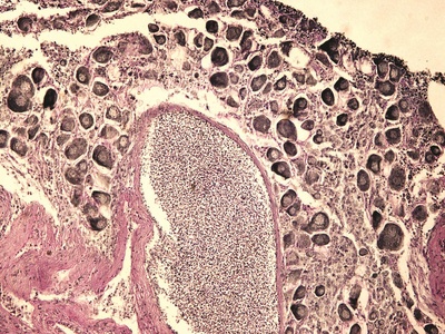
| Amygdala | Medial temporal lobe(limbic system) | Principal neurons,interneurons,astrocytes,oligodendrocytes | Emotional processing,fear learning,emotional memory modulation |
Images and Descriptions
Cerebral cortex
The cerebral cortex is the brain’s outer layer of neural tissue responsible for perception, decision-making, language, and voluntary movements. It contains specialized layers of neurons and supporting glia that integrate sensory information and guide complex behaviors and thought.

Cerebellar cortex
The cerebellar cortex is a tightly folded nervous tissue layer that refines movement, balance, and posture. Purkinje and granule neurons work with glial cells to coordinate timing and precision of muscle activity and adapt motor patterns through learning.
Hippocampus
The hippocampus is a curved structure critical for forming new memories and navigating space. Packed with excitatory neurons and glia, it helps encode experiences, consolidate short-term memories into long-term storage, and supports certain types of learning.
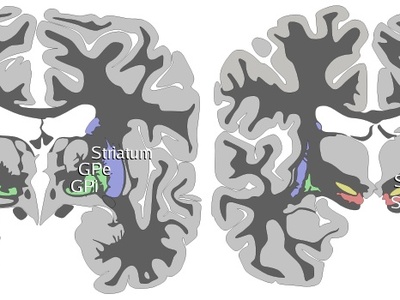
Basal ganglia (striatum)
The basal ganglia are groups of nuclei including the striatum that regulate voluntary movement, reward processing, and habit learning. Interconnected neurons and glia shape motor signals and suppress unwanted movements through inhibitory circuits.
Thalamus
The thalamus is a central relay station of nervous tissue that channels most sensory information to the cerebral cortex. Thalamic neurons and glia filter and coordinate signals, influencing attention, sleep-wake states, and aspects of motor control.
Hypothalamus
The hypothalamus is a small but vital neural region that maintains body balance—temperature, hunger, thirst, and circadian rhythms. Specialized neurons interact with glia to control hormone release and coordinate autonomic and behavioral responses to internal needs.

Brainstem
The brainstem is a compact assembly of nervous tissue connecting brain and spinal cord. It contains nuclei and pathways that regulate breathing, cardiovascular function, sleep-wake cycles, and many reflexes, relying on diverse neurons and supporting glial cells.
Spinal cord
The spinal cord is a cylindrical nervous tissue tract conveying sensory signals to the brain and motor commands to the body. Its organized gray and white matter contains neuronal cell bodies and glia that mediate reflexes and rapid sensorimotor processing.
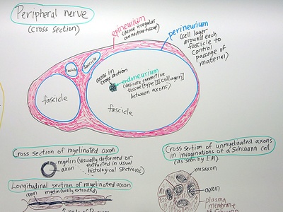
Peripheral nerve
Peripheral nerves are bundles of axons and Schwann cells that transmit sensory and motor information between the spinal cord or brain and body tissues. Encased by connective sheaths, they enable sensation, voluntary movement, and autonomic control across the body.
Dorsal root ganglion
Dorsal root ganglia are clusters of sensory neuron cell bodies located on spinal roots. These ganglia house primary afferent neurons and satellite glia, converting peripheral sensory inputs into electrical signals sent into the spinal cord for processing and reflexes.
Autonomic ganglia
Autonomic ganglia are groups of neurons that relay sympathetic or parasympathetic commands to target organs. Neurons and glia in these ganglia process and shape autonomic signals controlling heart rate, digestion, pupil size, and other involuntary functions.
Enteric nervous system (myenteric plexus)
The enteric nervous system is a dense network of neurons and glia embedded in the gut wall that independently controls intestinal movement, secretion, and blood flow. It integrates local sensory input and coordinates complex peristaltic and secretory behaviors.
Retina
The retina is a layered nervous tissue that captures light and begins vision processing. Neurons including photoreceptors, bipolar and ganglion cells, together with retinal glia, convert light into electrical signals sent along the optic nerve to the brain.
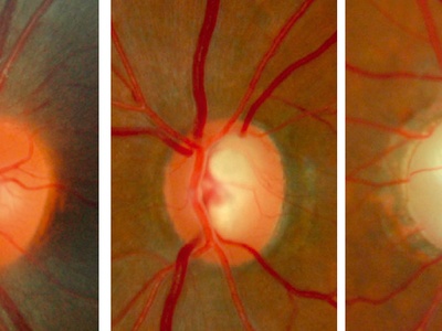
Optic nerve
The optic nerve is a CNS white-matter tract formed by retinal ganglion cell axons and myelinating oligodendrocytes. It conveys visual signals from the eye to central visual centers, and is bundled with supportive glia that maintain axonal function.
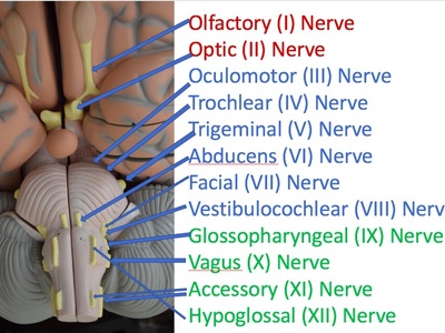
Cranial nerves
Cranial nerves are bundles of neuronal fibers connecting the brain to head, neck, and visceral structures. Composed mainly of axons and glia, they carry sensory information, control muscles, and regulate autonomic functions depending on the specific nerve.
Olfactory bulb
The olfactory bulb is a neural structure that receives input from olfactory receptor neurons and refines odor signals. Neurons and specialized glia organize odor maps, enhance contrast, and relay processed smell information to cortical areas for perception and memory associations.
Trigeminal ganglion
The trigeminal ganglion houses sensory neuron cell bodies transmitting touch, pain, and temperature from the face and mouth. Satellite glia support these neurons, which convey peripheral signals to brainstem nuclei for further processing and reflex actions.
Ventral horn (spinal motor nucleus)
The ventral horn contains motor neuron cell bodies that directly innervate skeletal muscles. Integrated with interneurons and glia, this nervous tissue generates voluntary and reflexive motor outputs, translating spinal and supraspinal signals into muscle contraction.
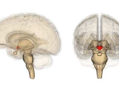
Substantia nigra
The substantia nigra is a midbrain nucleus whose dopaminergic neurons influence movement control and reward circuits. Loss of these neurons causes motor deficits; local glia support metabolic needs and influence neuronal health and signaling.
Amygdala
The amygdala is a cluster of nuclei involved in processing emotions and forming emotional memories. Neurons and glia in this nervous tissue detect salient stimuli, coordinate fear responses, and influence behaviors linked to reward and social interactions.