Across the nervous system, specialized cells take on roles from sensing the environment to driving movement and coordinating thought. Looking at specific neuron types helps connect microscopic structure to larger functions like reflexes, perception, and voluntary action.
There are 20 Examples of Neurons, ranging from the Alpha motor neuron to the Thalamocortical relay neuron. For each entry you’ll find below the data organized by Classification, Main function, Typical location.
How are different neuron types identified and named?
Neurons are classified by shape, connectivity, neurotransmitter, electrophysiological properties and role (sensory, motor, interneuron), so names often reflect one or more of those traits; for example, “Alpha motor neuron” denotes a large motor cell that innervates muscle fibers, while “Thalamocortical relay neuron” indicates its connection and function in relaying sensory information to cortex.
Which of these neuron examples are most relevant to disease or injury?
Many examples have clinical importance: motor neurons are central to ALS and spinal injuries, sensory and thalamic relay neurons relate to pain and sensory disorders, and specific interneurons are implicated in epilepsy and psychiatric conditions — knowing the cell type helps target research and treatment.
Examples of Neurons
| Name | Classification | Main function | Typical location |
|---|---|---|---|
| Pyramidal cell | Excitatory projection neuron | Excitatory output and integration; cortical long-range signaling | Cerebral cortex; hippocampus |
| Purkinje cell | Inhibitory projection neuron (cerebellar) | Inhibit cerebellar nuclei; coordinate motor timing and learning | Cerebellar cortex |
| Cerebellar granule cell | Excitatory interneuron (very small) | Relay mossy fiber input via parallel fibers to Purkinje layer | Cerebellar cortex granule layer |
| Golgi cell | Inhibitory interneuron (cerebellar) | Provide feedback inhibition to granule cells, shaping input timing | Cerebellar granule layer |
| Dentate granule cell | Excitatory projection neuron (hippocampal) | Support pattern separation and memory encoding | Dentate gyrus, hippocampus |
| Medium spiny neuron | Inhibitory projection neuron | Integrate cortical input and inhibit basal ganglia output pathways | Striatum (caudate nucleus, putamen) |
| Alpha motor neuron | Lower motor neuron; cholinergic projection | Drive skeletal muscle contraction and mediate reflexes | Ventral horn of spinal cord; brainstem motor nuclei |
| Dorsal root ganglion neuron | Primary sensory neuron | Transmit touch, temperature, pain, and proprioceptive signals to spinal cord | Dorsal root ganglia (spinal) |
| Nociceptor | Primary sensory pain neuron | Detect and signal damaging or potentially damaging stimuli (pain) | Dorsal root and trigeminal ganglia; peripheral nerves |
| Retinal ganglion cell | Output neuron (retina) | Transmit visual signals from retina to brain via optic nerve | Retina (ganglion cell layer) |
| Rod photoreceptor | Sensory receptor neuron (photoreceptor) | Detect low-light (scotopic) vision | Retina, peripheral retina |
| Cone photoreceptor | Sensory receptor neuron (photoreceptor) | Detect color and support high-acuity daylight vision | Retina, concentrated in fovea (primates) |
| Bipolar cell (retina) | Relay interneuron (retina) | Connect photoreceptors to ganglion cells, transmit visual signals | Retina (inner nuclear layer) |
| Amacrine cell | Retinal interneuron (diverse) | Modulate and integrate signals laterally in inner retina | Retina (inner nuclear layer/inner plexiform layer) |
| Horizontal cell | Retinal interneuron | Provide lateral inhibition to shape contrast and receptive fields | Retina (outer plexiform layer) |
| Mitral cell | Principal projection neuron (olfactory bulb) | Relay odor information from olfactory bulb to cortex | Olfactory bulb |
| Olfactory receptor neuron | Primary sensory neuron (chemosensory) | Detect odor molecules and transduce them into neural signals | Olfactory epithelium (nasal cavity) |
| Thalamocortical relay neuron | Excitatory thalamic projection neuron | Convey sensory and modulatory signals between thalamus and cortex | Thalamic nuclei (sensory and association) |
| Parvalbumin-positive interneuron | Fast-spiking inhibitory interneuron (molecular) | Provide fast, perisomatic inhibition to synchronize networks | Cortex, hippocampus, many brain regions |
| Chandelier cell | Morphologically unique inhibitory interneuron | Inhibit axon initial segments to powerfully regulate spike output | Cortex, hippocampus |
Images and Descriptions
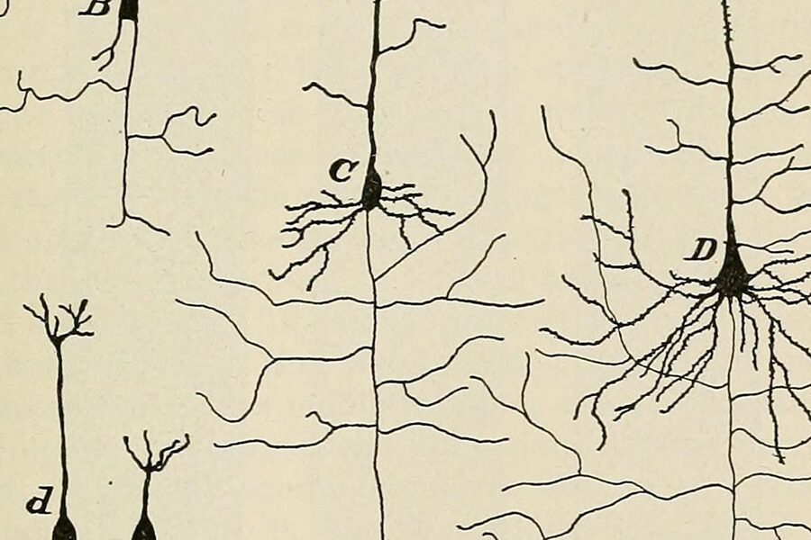
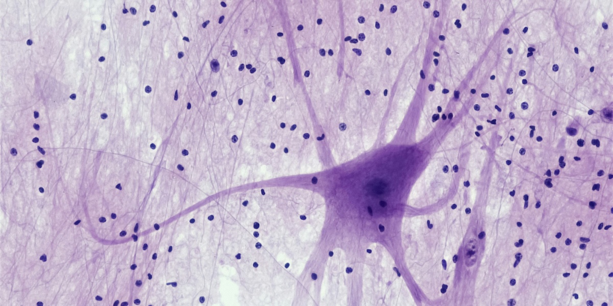
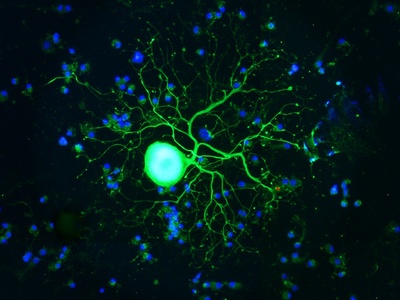
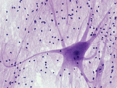
Pyramidal cell
Large excitatory neuron with a pyramid-shaped soma and prominent apical dendrite. Pyramidal cells integrate many inputs and send long-range projections across brain regions, supporting cognition, memory, and major cortical computations; diverse subtypes have specialized circuit roles.
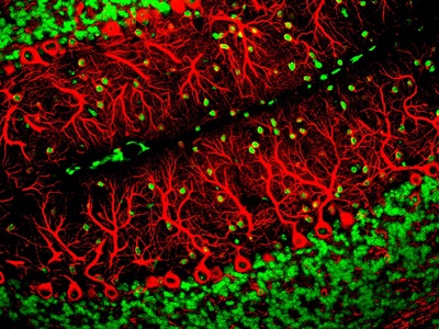
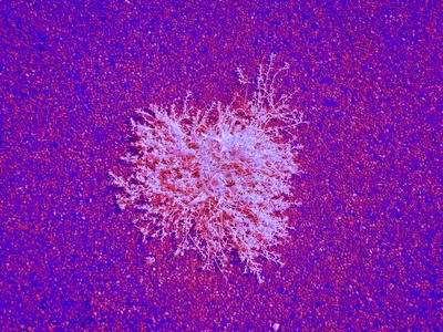
Purkinje cell
Huge neurons with an elaborate, planar dendritic tree and a single inhibitory output. Purkinje cells receive massive input, generate distinctive spike patterns, and refine motor coordination and timing by inhibiting deep cerebellar nuclei essential for smooth movement and motor learning.

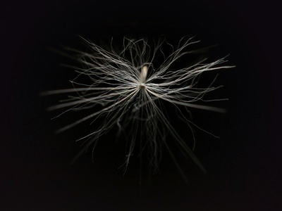
Cerebellar granule cell
Tiny, extremely numerous excitatory neurons that receive mossy fiber inputs and send parallel fibers across the cerebellar cortex. They are the most abundant neuron type and critically shape sensorimotor integration, timing, and cerebellar computations.
Golgi cell
GABAergic interneurons that regulate granule cell activity through feedforward and feedback inhibition. Golgi cells control timing and gain of cerebellar inputs, influencing precision of motor coordination and the cerebellum’s learning functions.
Dentate granule cell
Principal excitatory neuron of the dentate gyrus with compact dendrites and sparse firing. Dentate granule cells orthogonalize incoming signals to aid pattern separation during memory formation and are notable for adult neurogenesis in many species.
Medium spiny neuron
GABAergic projection neurons that form the bulk of the striatum. With densely spined dendrites, medium spiny neurons integrate cortical and dopaminergic signals to control movement, habit formation, and action selection within basal ganglia circuits.
Alpha motor neuron
Large cholinergic neurons whose axons innervate muscle fibers at neuromuscular junctions. Alpha motor neurons translate neural commands into muscle contraction, mediate reflexes, and are vulnerable in diseases like amyotrophic lateral sclerosis.
Dorsal root ganglion neuron
Cell bodies of peripheral sensory neurons that relay diverse sensations from the body to the spinal cord. DRG neurons have varied specializations for touch, temperature, pain, and proprioception and are essential for somatosensation and reflexes.
Nociceptor
Specialized sensory neurons that respond to high-threshold thermal, mechanical, or chemical stimuli. Nociceptors initiate pain signals, sensitize after injury, and drive protective reflexes and inflammatory responses; they are central targets for pain therapies.
Retinal ganglion cell
Diverse output neurons that integrate inputs from bipolar and amacrine cells and send visual information through the optic nerve. Different retinal ganglion cell types encode brightness, contrast, motion, and other features for downstream visual centers.
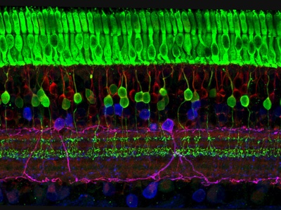
Rod photoreceptor
Light-sensitive photoreceptors specialized for dim-light vision. Rods have very high sensitivity with no color discrimination, converge onto bipolar cells, dominate peripheral retina, and enable night vision and navigation in near-dark conditions.

Cone photoreceptor
Photoreceptors specialized for bright light and color discrimination, often in three spectral types in primates. Cones provide high spatial acuity, support reading and color perception, and are concentrated in the fovea for detailed vision.
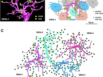
Bipolar cell (retina)
Vertical relay neurons receiving input from rods and cones and transmitting to ganglion cells. On and off bipolar types encode increases or decreases in light intensity, shaping early contrast and luminance information in the visual pathway.
Amacrine cell
A heterogeneous class of inhibitory and modulatory interneurons that shape temporal and spatial features of visual signals before ganglion cell output. Amacrine cells contribute to motion detection, contrast sensitivity, and complex visual computations.
Horizontal cell
Lateral inhibitory neurons connecting photoreceptors and bipolar cells to sharpen contrast and influence color processing. Horizontal cells help create center-surround receptive fields, enhancing edge detection and spatial contrast in vision.
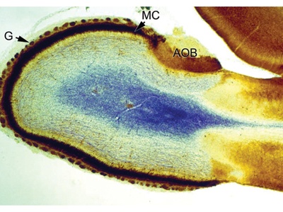
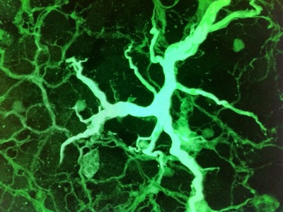
Mitral cell
Main output neurons of the olfactory bulb that receive input from receptor neuron glomeruli and transmit odor representations to olfactory cortex. Mitral cells integrate local circuits and shape odor tuning through lateral interactions.
Olfactory receptor neuron
Sensory neurons in the nose that express specific receptor families and send axons into the olfactory bulb. Each neuron type responds to particular odorants, creating the combinatorial code underlying smell perception.
Thalamocortical relay neuron
Excitatory thalamic neurons that transmit sensory signals and state-dependent rhythms to cortex, gating information flow. They operate in distinct firing modes to influence perception, attention, and sleep-wake dynamics.
Parvalbumin-positive interneuron
Molecularly defined fast-spiking GABAergic interneurons that target cell bodies and proximal dendrites to control timing and oscillations. PV interneurons are key for gamma rhythms, precise temporal control, and maintaining excitation–inhibition balance in circuits.
Chandelier cell
Distinctive interneurons with vertical arrays of axonal cartridges that synapse on axon initial segments of pyramidal cells. Chandelier cells exert powerful control over whether targets fire, strongly shaping network excitability and output.