In the human body, skeletal muscles form the visible and functional framework that lets you sit, reach, run and smile—they connect bones, shape movement and influence posture across every activity and environment. Whether you’re studying for a test, rehabbing an injury, or just curious about how muscles work, a clear list makes learning faster.
There are 25 Examples of Skeletal Muscles, ranging from Biceps brachii to Vastus lateralis; for each entry you’ll find below the Body region,Primary action,Innervation so you can compare location, function and nerve supply at a glance—useful for study, teaching or clinical reference, and you’ll find below.
How should I use this list to study muscle function effectively?
Use the table to link location with action and innervation: identify the Body region first, note the Primary action to visualize movement, then check the Innervation to understand clinical testing or injury patterns; quizzing yourself by naming the action or nerve for a given muscle improves recall.
Will this list help with clinical exams or anatomy labs?
Yes—having 25 representative muscles with region, action and nerve lets you practice palpation, manual muscle tests and nerve assessments efficiently; focus on commonly tested muscles like the Biceps brachii and Vastus lateralis while scanning the full list for regional patterns.
Examples of Skeletal Muscles
| Name | Body region | Primary action | Innervation |
|---|---|---|---|
| Biceps brachii | anterior arm | elbow flexion, forearm supination | musculocutaneous nerve |
| Triceps brachii | posterior arm | elbow extension | radial nerve |
| Deltoid | shoulder | shoulder abduction, flexion, extension | axillary nerve |
| Pectoralis major | anterior chest | shoulder flexion, adduction, internal rotation | medial and lateral pectoral nerves |
| Latissimus dorsi | back | shoulder extension, adduction, internal rotation | thoracodorsal nerve |
| Trapezius | upper back/neck | scapular elevation, retraction, rotation | spinal accessory nerve (CN XI), C3–C4 |
| Rectus abdominis | anterior abdomen | trunk flexion | thoracoabdominal nerves (T7–T11) and subcostal (T12) |
| External oblique | anterolateral abdomen | trunk rotation, lateral flexion | thoracoabdominal nerves (T7–T11) and subcostal (T12) |
| Gluteus maximus | buttock | hip extension, external rotation | inferior gluteal nerve |
| Gluteus medius | lateral hip | hip abduction, medial rotation | superior gluteal nerve |
| Rectus femoris | anterior thigh | knee extension, hip flexion | femoral nerve |
| Vastus lateralis | anterior thigh | knee extension | femoral nerve |
| Biceps femoris | posterior thigh | knee flexion, hip extension (long head) | sciatic nerve (tibial and common fibular parts) |
| Semitendinosus | posterior thigh | knee flexion, hip extension | tibial portion of sciatic nerve |
| Gastrocnemius | posterior calf | plantarflexion, knee flexion | tibial nerve |
| Soleus | posterior lower leg | plantarflexion | tibial nerve |
| Tibialis anterior | anterior lower leg | ankle dorsiflexion, inversion | deep peroneal (fibular) nerve |
| Masseter | jaw | jaw elevation (chewing) | mandibular branch of trigeminal nerve (V3) |
| Temporalis | temple | jaw elevation, retraction | mandibular nerve (V3) |
| Sternocleidomastoid | neck | head rotation, neck flexion, assists breathing | spinal accessory nerve (CN XI), C2–C3 |
| Iliocostalis | back | spine extension, lateral flexion | dorsal rami of spinal nerves |
| Brachialis | anterior arm | elbow flexion | musculocutaneous nerve; some radial fibers |
| Brachioradialis | forearm | elbow flexion (mid-position) | radial nerve |
| Pronator teres | forearm | forearm pronation, assists elbow flexion | median nerve |
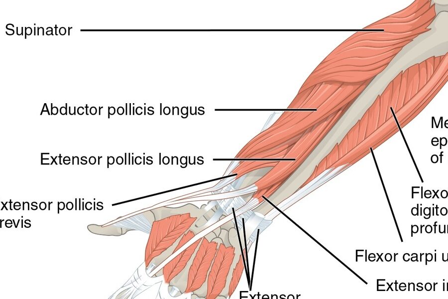
| Supinator | forearm | forearm supination | deep branch of radial nerve (posterior interosseous) |
Images and Descriptions
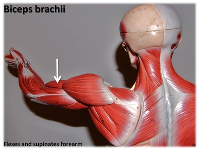
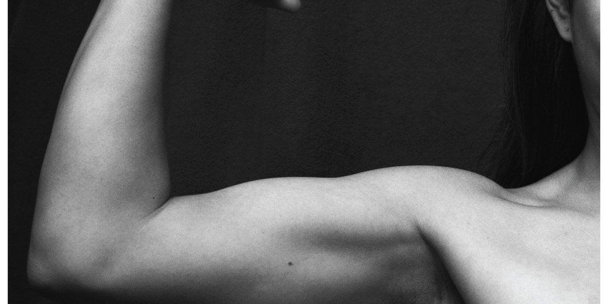
Biceps brachii
A two-headed muscle on the front of the upper arm. It bends the elbow and turns the palm upward (supination). Commonly felt when you flex; important for lifting, carrying, and many daily tasks. Injuries cause weakness in arm flexion.
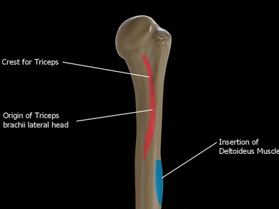
Triceps brachii
Large three-headed muscle on the back of the upper arm responsible for straightening the elbow. Key for pushing motions like push-ups and pushing doors. Strains or tendon injuries reduce arm extension strength and make pushing tasks difficult.
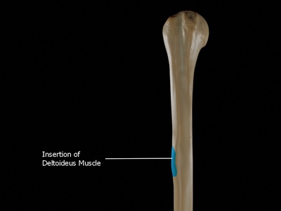
Deltoid
Triangular shoulder muscle that lifts the arm away from the body and assists front and back arm movements. It gives the shoulder its rounded shape. Injuries limit overhead motion and reduce shoulder strength during daily activities and sports.
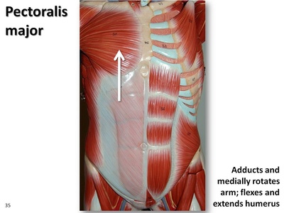
Pectoralis major
Broad chest muscle that brings the arm across the body, helps lift the arm forward, and rotates it inward. Important for pushing, hugging, and lifting. Strains can limit chest and shoulder activities and occur with heavy lifting.
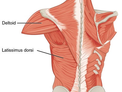
Latissimus dorsi
Large flat back muscle that pulls the arm down and back and rotates it inward. Used in climbing, swimming, and pulling motions. Strength and flexibility affect posture and shoulder health; commonly targeted in back workouts.
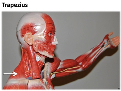
Trapezius
Large upper back and neck muscle that moves and stabilizes the shoulder blades: elevates, retracts, and rotates them. Important for posture, carrying loads, and head support. Tension here often causes neck and upper back pain.
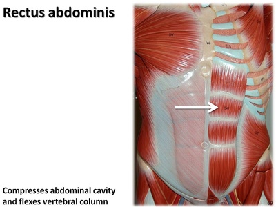
Rectus abdominis
Flat paired muscle running down the front of the abdomen — the “six-pack.” It flexes the spine, stabilizes the trunk during movement, and supports posture and breathing. Core strength reduces back pain and improves balance.
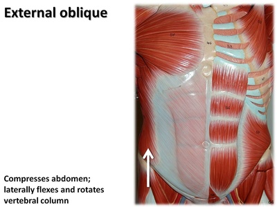
External oblique
Broad superficial abdominal muscle on the sides that helps rotate and bend the trunk and compress the abdomen. It supports posture and breathing and plays a role in twisting motions like reaching or throwing.
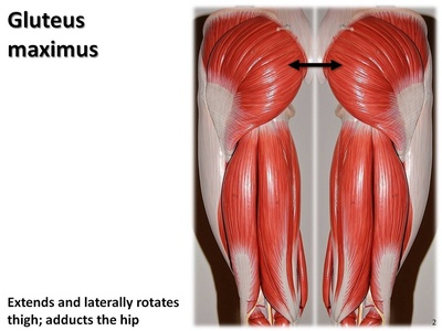
Gluteus maximus
Large buttock muscle that extends and laterally rotates the hip, powering activities such as climbing stairs, standing from a chair, and sprinting. Strong glutes support posture and lower back health; weakness contributes to hip and knee problems.
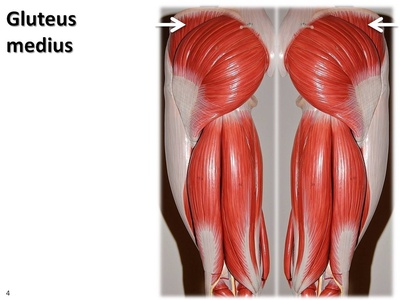
Gluteus medius
Located on the outer hip, this muscle abducts and medially rotates the thigh, stabilizing the pelvis during walking and single-leg stance. Weakness can cause hip pain and an unstable gait; crucial for balance and efficient walking.
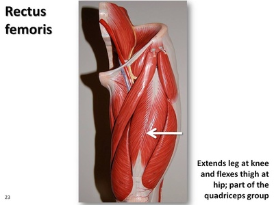
Rectus femoris
One of the four quadriceps on the front of the thigh. It straightens the knee and assists hip flexion. Active in walking, running, and kicking; strains are common in athletes and cause front-thigh pain and reduced leg power.
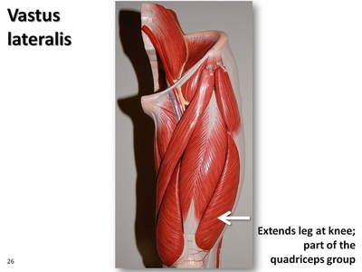
Vastus lateralis
Lateral quadriceps muscle that extends the knee and contributes to walking, running, and standing up. It’s a common injection site and often trained for knee stability and athletic performance.
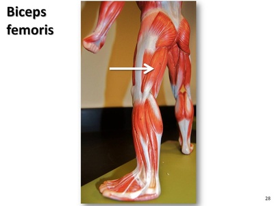
Biceps femoris
Part of the hamstrings at the back of the thigh with long and short heads. It bends the knee and assists hip extension (long head). Hamstring strains often involve this muscle and affect sprinting and sudden acceleration.
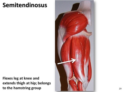
Semitendinosus
Middle hamstring muscle behind the thigh that flexes the knee and extends the hip. Important for running and posture; tears or tightness can cause posterior thigh pain and limit athletic movements.
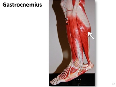
Gastrocnemius
Large calf muscle with two heads that plantarflexes the ankle (pushes the foot down) and flexes the knee. Prominent when standing on tiptoe, it’s crucial for jumping and walking; strains cause calf pain and difficulty with push-off.
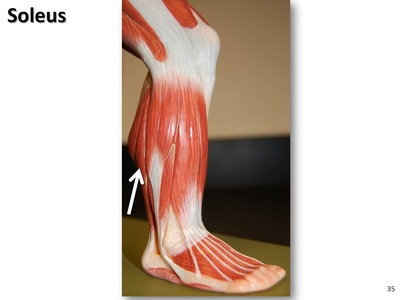
Soleus
Deep calf muscle beneath the gastrocnemius that primarily plantarflexes the ankle and helps maintain posture during standing and walking. It works constantly for balance and endurance activities; tightness can limit ankle motion and mobility.
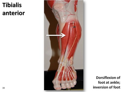
Tibialis anterior
Front lower-leg muscle that lifts the foot (dorsiflexion) and inverts the ankle. It prevents toes from dragging during walking and often becomes sore after long uphill walking or when new to running.
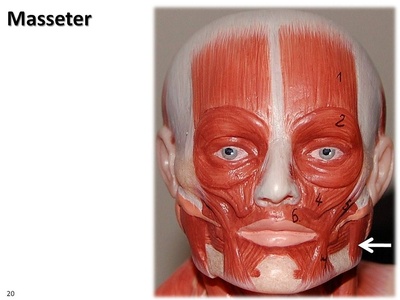
Masseter
Powerful jaw muscle on the side of the face that elevates the mandible for chewing and clenching. It shapes the jaw and can become overactive in teeth grinding or TMJ disorders, causing pain and headaches.
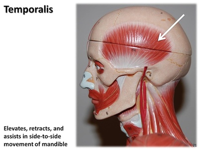
Temporalis
Fan-shaped muscle on the temple that elevates and retracts the jaw during chewing. It works with the masseter for bite force; tension here can contribute to headaches and jaw pain in bruxism or TMJ problems.
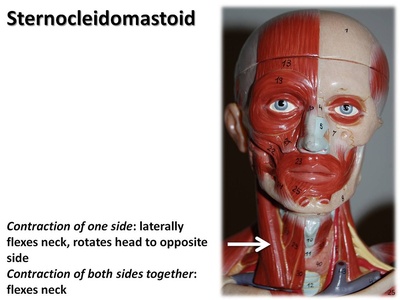
Sternocleidomastoid
Large paired neck muscle that turns the head to the opposite side and flexes the neck. It also helps raise the sternum during deep breathing. Tightness causes neck pain, headaches, and limited movement.

Iliocostalis
A lateral component of the erector spinae group that extends and laterally bends the spine, helping maintain upright posture. It supports lifting and twisting movements; weakness or spasm contributes to lower back pain and stiffness.
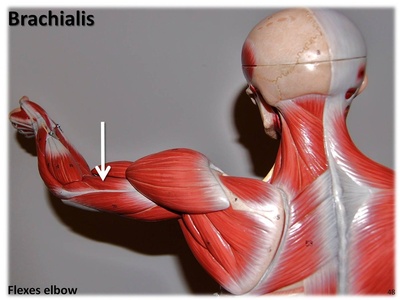
Brachialis
Deep anterior arm muscle under the biceps that is a primary elbow flexor in all forearm positions. Important for lifting and pulling; injury leads to difficulty bending the elbow and reduced arm strength.
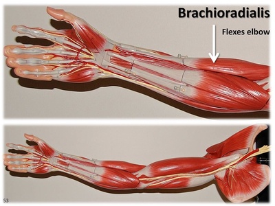
Brachioradialis
Forearm muscle visible near the elbow that flexes the elbow, especially with the forearm in a mid-pronated position. Useful for hammering motions and everyday lifting; commonly engaged during rowing or curling movements.
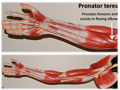
Pronator teres
Forearm muscle that turns the palm downward (pronation) and assists elbow flexion. It helps activities like turning a doorknob; compression of the median nerve near this muscle can cause forearm pain or numbness.
Supinator
Deep forearm muscle that rotates the forearm to turn the palm upward (supination), working with the biceps. Important for screwdriver-like motions; injury or nerve entrapment here can weaken supination, affecting many daily tasks.