From the heart out to the smallest peripheral branches, the arterial system carries oxygenated blood to every organ and tissue. Tracing those vessels helps students, clinicians and curious readers understand where blood comes from and which regions depend on each branch.
There are 85 Arteries, ranging from Abdominal aorta to Vertebrobasilar (Basilar) artery. For each entry we list Origin,Territory supplied,Typical diameter (mm) so you can compare sources, targets and size — you’ll find below.
How is “Typical diameter (mm)” determined and why does it matter?
Typical diameters come from anatomical studies, imaging (CT, ultrasound, angiography) and averaged measurements; they give a practical sense of vessel size for understanding flow, choosing catheter sizes, or assessing risk (for example, aneurysm thresholds). Use them as general guides but expect individual variation.
Will this list tell me which artery supplies a specific organ?
Yes — the Territory supplied column links arteries to the regions or organs they perfuse, making the list a quick reference for anatomy or clinical correlation. Keep in mind anatomical variants exist, so consult imaging or surgical notes for patient-specific planning.
Arteries
| Name | Origin | Territory supplied | Typical diameter (mm) |
|---|---|---|---|
| Ascending aorta | Left ventricle | Heart and systemic circulation origin | 30 |
| Aortic arch | Ascending aorta | Head, neck, upper limbs via branches | 25 |
| Thoracic aorta | Aortic arch | Chest wall, thoracic organs, intercostal spaces | 25 |
| Abdominal aorta | Diaphragmatic aorta | Abdominal organs and lower limbs via branches | 20 |
| Brachiocephalic trunk | Aortic arch | Right head, neck, right upper limb | 10 |
| Common carotid artery | Brachiocephalic (R)/Aortic arch (L) | Head and neck via internal/external branches | 7 |
| Subclavian artery | Brachiocephalic (R)/Aortic arch (L) | Upper limb and posterior brain via vertebral artery | 9 |
| Vertebral artery | Subclavian artery | Posterior brain, cerebellum, brainstem | 4 |
| Internal carotid artery | Common carotid artery | Anterior brain, eyes, forehead | 6 |
| External carotid artery | Common carotid artery | Face, scalp, neck, meninges | 5 |
| Vertebrobasilar (Basilar) artery | Vertebral arteries (fusion) | Brainstem, cerebellum, posterior cerebrum | 3 |
| Middle cerebral artery (MCA) | Internal carotid artery | Lateral cerebral hemispheres, motor and speech areas | 3 |
| Anterior cerebral artery (ACA) | Internal carotid artery | Medial frontal and parietal lobes | 2 |
| Posterior cerebral artery (PCA) | Basilar artery | Occipital lobes, inferior temporal lobes | 2 |
| Anterior communicating artery | Anterior cerebral arteries | Connects ACAs within circle of Willis | 2 |
| Posterior communicating artery | Internal carotid/basilar system | Connects anterior and posterior circulations | 1.5 |
| Ophthalmic artery | Internal carotid artery | Eye, orbit, lacrimal gland | 2 |
| Superior thyroid artery | External carotid artery | Thyroid gland, larynx, neck tissues | 2 |
| Lingual artery | External carotid artery | Tongue, floor of mouth | 2 |
| Facial artery | External carotid artery | Face, lips, nose, submandibular region | 2 |
| Occipital artery | External carotid artery | Posterior scalp and neck muscles | 1.5 |
| Maxillary artery | External carotid artery | Deep face, nasal cavity, palate, teeth | 2.5 |
| Superficial temporal artery | External carotid artery | Temporal scalp, auricular region | 2 |
| Internal thoracic artery | Subclavian artery | Anterior chest wall, breasts | 3 |
| Intercostal arteries | Thoracic aorta | Intercostal muscles, spinal cord via radicular branches | 2 |
| Celiac trunk | Abdominal aorta | Stomach, spleen, liver, pancreas via three branches | 8 |
| Common hepatic artery | Celiac trunk | Liver, stomach, pancreas, duodenum | 5 |
| Hepatic artery proper | Common hepatic artery | Liver, gallbladder | 4 |
| Gastroduodenal artery | Common hepatic artery | Stomach, duodenum, pancreas | 3 |
| Right gastroepiploic (gastro-omental) artery | Gastroduodenal artery | Greater curvature of stomach | 3 |
| Left gastroepiploic (gastro-omental) artery | Splenic artery | Greater curvature of stomach | 3 |
| Short gastric arteries | Splenic artery | Fundus of stomach | 2 |
| Splenic artery | Celiac trunk | Spleen, pancreatic tail, stomach | 5 |
| Superior mesenteric artery (SMA) | Abdominal aorta | Small intestine, proximal colon, pancreas | 7 |
| Inferior pancreaticoduodenal artery | SMA | Pancreas and duodenum | 2 |
| Inferior mesenteric artery (IMA) | Abdominal aorta | Distal colon, rectum (hindgut) | 4 |
| Renal artery | Abdominal aorta | Kidney | 5 |
| Middle suprarenal artery | Abdominal aorta | Adrenal gland (suprarenal) | 2 |
| Inferior phrenic artery | Abdominal aorta/common trunk | Diaphragm, suprarenal glands | 2 |
| Common iliac artery | Abdominal aorta | Pelvis and lower limb via internal/external branches | 10 |
| External iliac artery | Common iliac artery | Lower limb (continues as femoral) | 7 |
| Internal iliac artery | Common iliac artery | Pelvis, gluteal region, perineum | 6 |
| Obturator artery | Internal iliac artery | Medial thigh, hip joint | 2 |
| Internal pudendal artery | Internal iliac artery | Perineum, external genitalia | 3 |
| Uterine artery | Internal iliac artery | Uterus, upper vagina | 3 |
| Ovarian artery | Abdominal aorta | Ovary, fallopian tube | 2.5 |
| Testicular (gonadal) artery | Abdominal aorta | Testis, epididymis | 2 |
| Femoral artery | External iliac artery | Thigh and lower limb via branches | 7 |
| Profunda femoris (deep femoral) artery | Femoral artery | Deep thigh muscles and femoral head collaterals | 4 |
| Popliteal artery | Femoral artery | Knee joint, lower leg via tibial branches | 6 |
| Anterior tibial artery | Popliteal artery | Anterior compartment of leg, dorsum of foot | 3 |
| Posterior tibial artery | Popliteal artery | Posterior compartment of leg, plantar foot | 4 |
| Peroneal (fibular) artery | Posterior tibial or tibial trunk | Lateral compartment of leg, fibula | 3 |
| Dorsalis pedis artery | Anterior tibial artery | Dorsum of foot, toes | 2 |
| Internal mammary artery | Internal thoracic artery | Anterior chest wall, breasts | 3 |
| Axillary artery | Subclavian artery | Shoulder, lateral thorax, upper limb | 6 |
| Brachial artery | Axillary artery | Arm, forearm via radial/ulnar branches | 5 |
| Radial artery | Brachial artery | Lateral forearm, hand (thenar eminence) | 2 |
| Ulnar artery | Brachial artery | Medial forearm, hand (palmar arches) | 2 |
| Profunda brachii (deep brachial) artery | Brachial artery | Posterior arm, triceps | 2 |
| Pulmonary artery (main) | Right ventricle | Pulmonary arteries to both lungs | 25 |
| Right pulmonary artery | Pulmonary trunk | Right lung | 15 |
| Left pulmonary artery | Pulmonary trunk | Left lung | 15 |
| Left coronary artery (LCA) | Left coronary cusp of aorta | Left heart, septum via branches | 4 |
| Left anterior descending artery (LAD) | Left coronary artery | Anterior interventricular septum and anterior LV | 3 |
| Left circumflex artery (LCx) | Left coronary artery | Lateral and posterior left ventricle | 3 |
| Right coronary artery (RCA) | Right coronary cusp of aorta | Right heart, inferior LV, AV node | 3 |
| Posterior descending artery (PDA) | RCA or LCx (variant) | Inferior interventricular septum, inferior LV wall | 2.5 |
| Superior gluteal artery | Internal iliac artery | Gluteal muscles, hip joint | 3 |
| Inferior gluteal artery | Internal iliac artery | Gluteus maximus, posterior thigh | 3 |
| Common palmar digital arteries | Superficial palmar arch | Fingers | 1 |
| Renal segmental arteries | Renal artery | Renal segments (kidney poles/areas) | 3 |
| Lumbar arteries | Abdominal aorta | Posterior abdominal wall, spinal cord branches | 2 |
| Median sacral artery | Abdominal aorta | Sacrum, coccyx, caudal lumbar vertebrae | 1.5 |
| Superior rectal artery | Inferior mesenteric artery | Upper rectum | 3 |
| Inferior epigastric artery | External iliac artery | Anterior abdominal wall | 3 |
| Superior pancreaticoduodenal artery | Gastroduodenal artery | Pancreas head, duodenum | 2 |
| Right colic artery | Superior mesenteric artery | Ascending colon | 2 |
| Middle colic artery | Superior mesenteric artery | Transverse colon | 2.5 |
| Ileocolic artery | Superior mesenteric artery | Ileum, cecum, appendix | 2 |
| Left colic artery | Inferior mesenteric artery | Descending colon | 2 |
| Internal maxillary artery | Maxillary artery | Deep facial structures, nasal cavity, palate | 2 |
| Posterior auricular artery | External carotid artery | Scalp behind ear, auricle | 1 |
| Superior laryngeal artery | Superior thyroid artery | Larynx | 1 |
| External pudendal artery | Femoral artery | Skin of scrotum/labia and lower abdomen | 1.5 |
Images and Descriptions
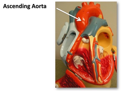
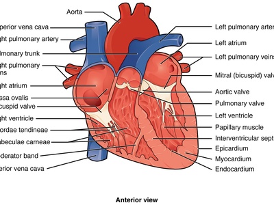
Ascending aorta
The initial upward segment of the aorta arising from the left ventricle; gives coronary arteries and supplies systemic circulation. Central conduit; aneurysm and dissection here are life-threatening and common surgically relevant conditions.

Aortic arch
Curved portion between ascending and descending aorta that gives rise to brachiocephalic trunk, left common carotid and left subclavian. Important in trauma, aortic disease and as landmark in vascular surgery and endovascular repair.
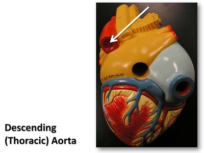
Thoracic aorta
Descending thoracic aorta supplies posterior mediastinum, intercostal arteries, esophagus and bronchi. Aortic aneurysms, traumatic transection and intercostal bleeding are clinically significant in thoracic vascular care.
Abdominal aorta
Continuation of thoracic aorta that gives celiac trunk, mesenteric and renal arteries and common iliacs. Major site of atherosclerosis and abdominal aortic aneurysms; key artery for visceral perfusion.

Brachiocephalic trunk
First major branch of the aortic arch on the right; divides into right common carotid and right subclavian arteries. Important in central vascular procedures and stroke/embolism pathways.
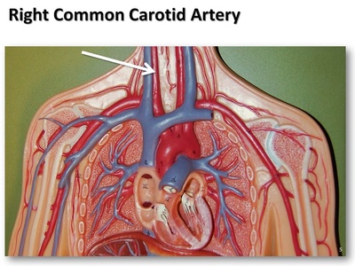
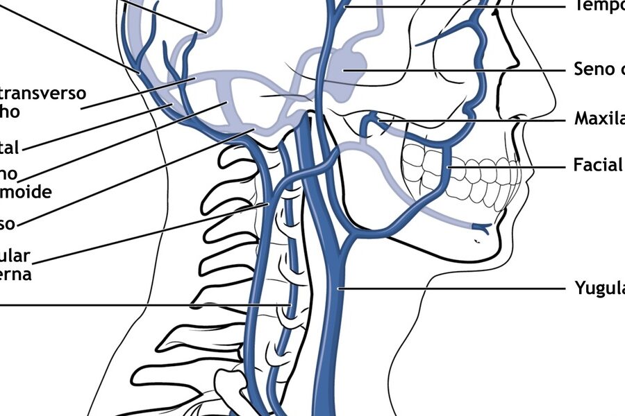
Common carotid artery
Main paired neck vessels that bifurcate into internal and external carotid arteries. Palpable pulses, carotid atherosclerosis causes stroke risk; carotid endarterectomy treats symptomatic stenosis.
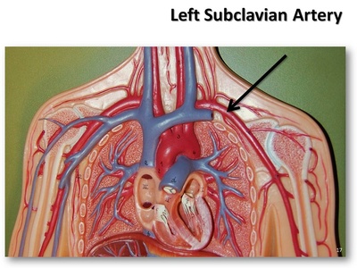
Subclavian artery
Supplies the upper limb and gives the vertebral artery to posterior brain and internal thoracic branches. Subclavian stenosis causes arm claudication and cerebral hypoperfusion; important in thoracic outlet syndrome.
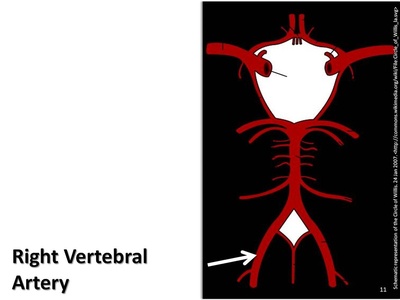
Vertebral artery
Ascends through cervical transverse foramina to join as basilar artery. Supplies posterior cerebral circulation; vertebral dissection causes posterior stroke and neck trauma can injure it.
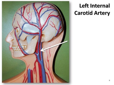
Internal carotid artery
Major intracranial artery entering skull via carotid canal; supplies anterior circulation (ICA branches, MCA, ACA). Atherosclerotic plaque causes ischemic stroke; carotid interventions target proximal disease.
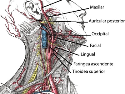
External carotid artery
Supplies extracranial structures including face, scalp, thyroid and oral cavity via multiple branches. Important for head and neck surgery and embolization in epistaxis or tumor bleeding.
Vertebrobasilar (Basilar) artery
Formed by vertebral arteries joining at the pons to form the basilar artery. Supplies brainstem and cerebellum; basilar occlusion causes severe stroke with high morbidity and mortality.

Middle cerebral artery (MCA)
Largest branch of ICA supplying lateral frontal, parietal and temporal lobes; territory of common ischemic strokes producing hemiparesis, aphasia and sensory loss. A key target in stroke therapy.
Anterior cerebral artery (ACA)
Supplies medial surfaces of frontal lobes and corpus callosum. ACA infarcts cause leg-predominant weakness and behavioral changes; anterior communicating aneurysms are common.

Posterior cerebral artery (PCA)
Supplies occipital cortex (vision) and medial/inferior temporal lobes. PCA strokes cause visual field defects; aneurysms and vascular malformations here affect visual processing.
Anterior communicating artery
Short midline connector between ACAs; common site for saccular (berry) aneurysms. Rupture leads to subarachnoid hemorrhage with high clinical consequence.
Posterior communicating artery
Links internal carotid and posterior cerebral arteries within the Circle of Willis. Important collateral route in carotid disease; site of aneurysms causing cranial nerve palsies.
Ophthalmic artery
First intracranial branch of the ICA supplying the retina and orbital structures. Central retinal artery occlusion causes sudden monocular blindness; ophthalmic artery injury risks vision loss.
Superior thyroid artery
First branch of external carotid supplying thyroid, larynx and anterior neck. Important in thyroid surgery and can be a source of hemorrhage or be ligated during procedures.

Lingual artery
Supplies the tongue and floor of mouth. Relevance in oral surgery, bleeding control and reconstructive flaps; lesions can affect taste and swallowing.
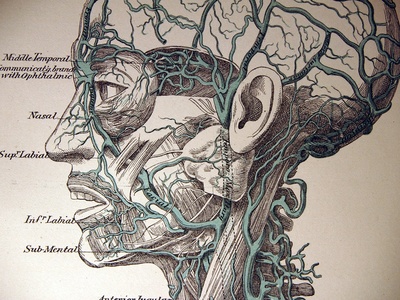
Facial artery
Tortuous artery supplying major facial structures including lips and nasal ala. Important for facial trauma, reconstructive surgery and epistaxis management.
Occipital artery
Supplies posterior scalp and deep neck muscles; palpable pulses occasionally. Involved in occipital neuralgia procedures and scalp flap surgeries.
Maxillary artery
Large terminal branch of external carotid supplying deep facial structures, nasal cavity and maxilla. Important in epistaxis, dental bleeding and embolization for trauma or tumors.

Superficial temporal artery
Terminal branch of external carotid supplying lateral scalp. Palpable pulse used in temporal arteritis diagnosis; biopsy of temporal artery assesses giant cell arteritis.
Internal thoracic artery
Descends behind sternum supplying anterior chest wall and pericardium. Commonly used as a graft in coronary artery bypass due to excellent patency; vulnerable in thoracic surgery.
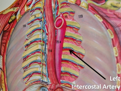
Intercostal arteries
Paired segmental arteries from thoracic aorta supplying ribs, chest wall and spinal cord radicular branches. Injury can cause hemothorax; radicular supply important in spinal cord ischemia risk.
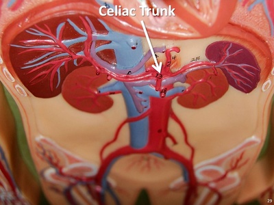
Celiac trunk
Short unpaired trunk dividing into left gastric, splenic and common hepatic arteries. Central for foregut perfusion; celiac disease or aneurysm has significant clinical implications.

Common hepatic artery
Branch of celiac trunk giving hepatic artery proper and gastroduodenal artery; supplies liver and upper GI. Variants affect transplant and hepatobiliary surgery planning.
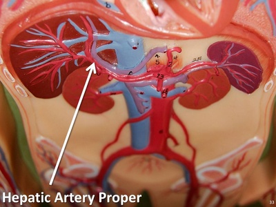
Hepatic artery proper
Terminal hepatic branch supplying arterial blood to liver and gallbladder alongside portal venous flow. Important in liver surgery, transplantation and trauma; variations alter surgical approach.
Gastroduodenal artery
Descends posterior to the stomach supplying the pylorus, duodenum and pancreas (gives superior pancreaticoduodenal). Common source of upper GI bleeding and target for embolization.

Right gastroepiploic (gastro-omental) artery
Runs along greater curvature supplying gastric antrum; used in some gastric and reconstructive surgeries. Important in bleeding peptic ulcer disease and gastric artery embolization.

Left gastroepiploic (gastro-omental) artery
Branch of splenic artery supplying greater curvature and omentum; used in gastric surgery and flap reconstruction. Vulnerable in splenic artery aneurysms or trauma.
Short gastric arteries
Small branches supplying the gastric fundus from the splenic artery. Clinically relevant in splenectomy and gastric surgery where they are ligated to prevent bleeding.
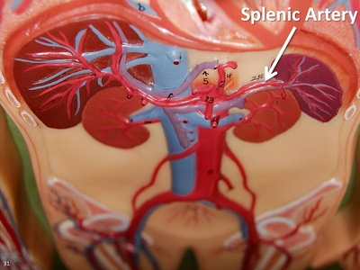
Splenic artery
Tortuous vessel supplying spleen and portions of pancreas and stomach. Prone to aneurysm; embolization or ligation performed in splenic trauma or hypersplenism.
Superior mesenteric artery (SMA)
Major midgut artery supplying jejunum, ileum, cecum, ascending and transverse colon. Acute SMA occlusion causes catastrophic intestinal ischemia; chronic mesenteric ischemia leads to postprandial pain and weight loss.
Inferior pancreaticoduodenal artery
Connects SMA to pancreaticoduodenal arcades supplying pancreas head and duodenum. Important collateral with superior pancreaticoduodenal artery; involved in pancreatitis-related bleeding.

Inferior mesenteric artery (IMA)
Supplies distal transverse colon, descending/sigmoid colon and upper rectum. IMA occlusion causes ischemic colitis; knowledge of collateral arcades matters in colorectal surgery.
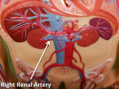
Renal artery
Paired arteries from abdominal aorta entering kidney hilum; essential for renal perfusion and blood pressure regulation. Renal artery stenosis causes renovascular hypertension; interventions include stenting and bypass.
Middle suprarenal artery
Small paired arteries from the aorta supplying adrenal cortex. Important in adrenal surgery and tumor vascularity; typically one of several adrenal arterial sources.
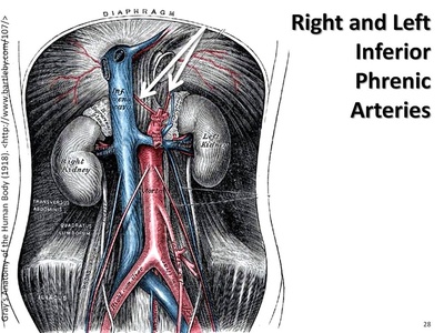
Inferior phrenic artery
Supplies diaphragm and gives branches to the adrenal glands and esophagus. Relevant in upper abdominal bleeding and hepatocellular carcinoma arterial supply.
Common iliac artery
Bifurcation of abdominal aorta forming internal and external iliac arteries. Site for atherosclerotic disease and endovascular repair; supplies pelvic organs and lower limb.
External iliac artery
Travels along pelvic brim to become femoral artery; supplies lower limb and gives deep circumflex and inferior epigastric branches. Important access site for endovascular procedures.
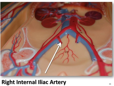
Internal iliac artery
Supplies pelvic organs, perineum and gluteal region via anterior and posterior divisions. Embolization used for pelvic hemorrhage and obstetric bleeding control.
Obturator artery
Runs along pelvic wall to supply medial thigh muscles and hip structures. Important in pelvic fractures and surgical repair due to anastomoses with femoral system.
Internal pudendal artery
Major artery to perineum and external genital organs; critical in urological and gynecologic surgery. Injury can cause severe pelvic hemorrhage; used in reconstructive procedures.
Uterine artery
Supplies uterus and has important role in pregnancy; uterine artery embolization treats fibroids and controls postpartum hemorrhage. Ligation considered in obstetric bleeding.
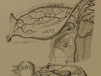
Ovarian artery
Direct branch from aorta supplying ovary and tube. Important in ovarian torsion and gynecological surgery; ovarian blood supply has dual arterial sources.
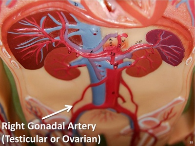
Testicular (gonadal) artery
Paired arteries from aorta supplying testes and scrotal contents. Clinical relevance in varicocele, torsion evaluation and during retroperitoneal surgery to avoid ischemic injury.
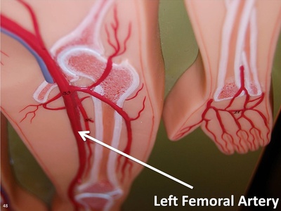
Femoral artery
Continuation of external iliac in the thigh; major access site for catheterization. Supplies lower limb; femoral pulse and occlusion are central to vascular assessment and interventions.
Profunda femoris (deep femoral) artery
Primary deep branch of femoral artery supplying thigh muscles and perforating branches. Important in supplying femoral head and for collateral flow in peripheral arterial disease.
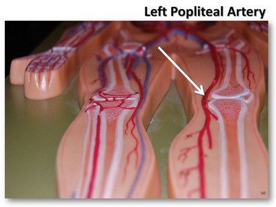
Popliteal artery
Continuation of femoral artery through adductor hiatus behind the knee. Bifurcates into anterior and posterior tibial arteries; popliteal aneurysms risk thrombosis and embolism.
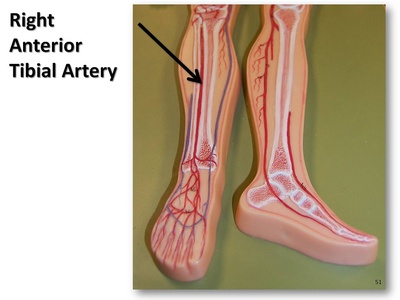
Anterior tibial artery
Passes to the anterior leg and continues as dorsalis pedis supplying dorsum of foot. Vulnerable in trauma; dorsalis pedis pulse assesses distal perfusion.
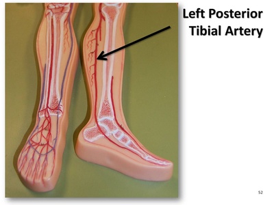
Posterior tibial artery
Supplies calf muscles and plantar foot via medial/lateral plantar arteries. Posterior tibial pulse used clinically; occlusion causes critical limb ischemia.
Peroneal (fibular) artery
Supplies lateral leg and fibula; perforating branches contribute to ankle and foot. Important collateral in tibial occlusive disease and in flap surgeries.
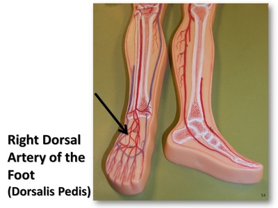
Dorsalis pedis artery
Continuation of anterior tibial on the dorsum of foot; palpable pulse used to assess distal perfusion. Occlusion indicates peripheral arterial disease risk.
Internal mammary artery
Alternate name for internal thoracic; commonly used in coronary bypass grafting due to long-term patency. Supplies anterior chest wall and breasts; preserved in thoracic surgery for grafting.
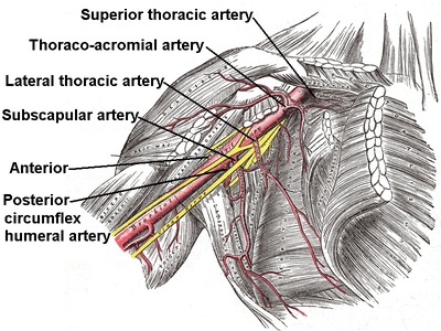
Axillary artery
Continuation of subclavian in the axilla supplying shoulder and upper limb via multiple branches. Important in trauma and regional anesthesia; access point in some vascular procedures.
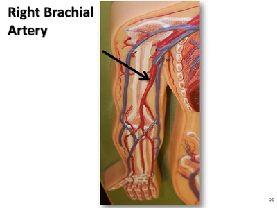
Brachial artery
Main artery of the upper arm continuing into forearm; palpable at antecubital fossa. Common site for blood pressure measurement, arterial lines and traumatic injury.
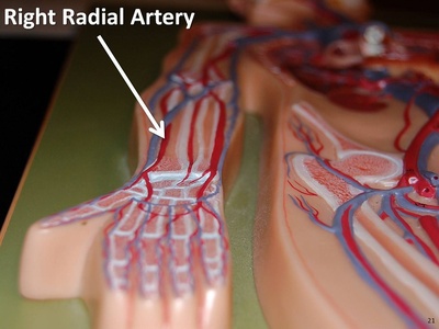
Radial artery
Supplies lateral forearm and hand; used for arterial blood gas sampling and coronary catheterization access (radial approach). Pulse felt at wrist; important in hand perfusion.
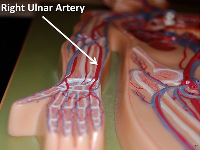
Ulnar artery
Supplies medial forearm and contributes majorly to superficial palmar arch. Dominant ulnar flow protects hand when radial artery compromised; target in bypass grafting.
Profunda brachii (deep brachial) artery
Deep branch of brachial artery supplying posterior arm musculature and humeral periosteum. Important in humeral fracture hemorrhage and surgical exposure of the arm.
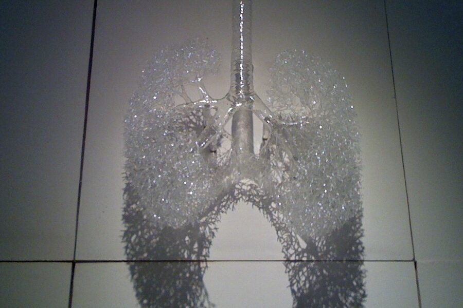
Pulmonary artery (main)
Carries deoxygenated blood from right ventricle to the lungs; bifurcates into right and left pulmonary arteries. Pulmonary embolism in this vessel is life-threatening; pressures measured in cardiology.
Right pulmonary artery
Branches from main pulmonary artery to supply right lung lobes. Embolism here causes right heart strain; anatomy relevant in pulmonary surgeries and interventions.
Left pulmonary artery
Supplies left lung lobes; courses anterior to descending aorta. Important in pulmonary embolism distribution and in thoracic surgical planning.
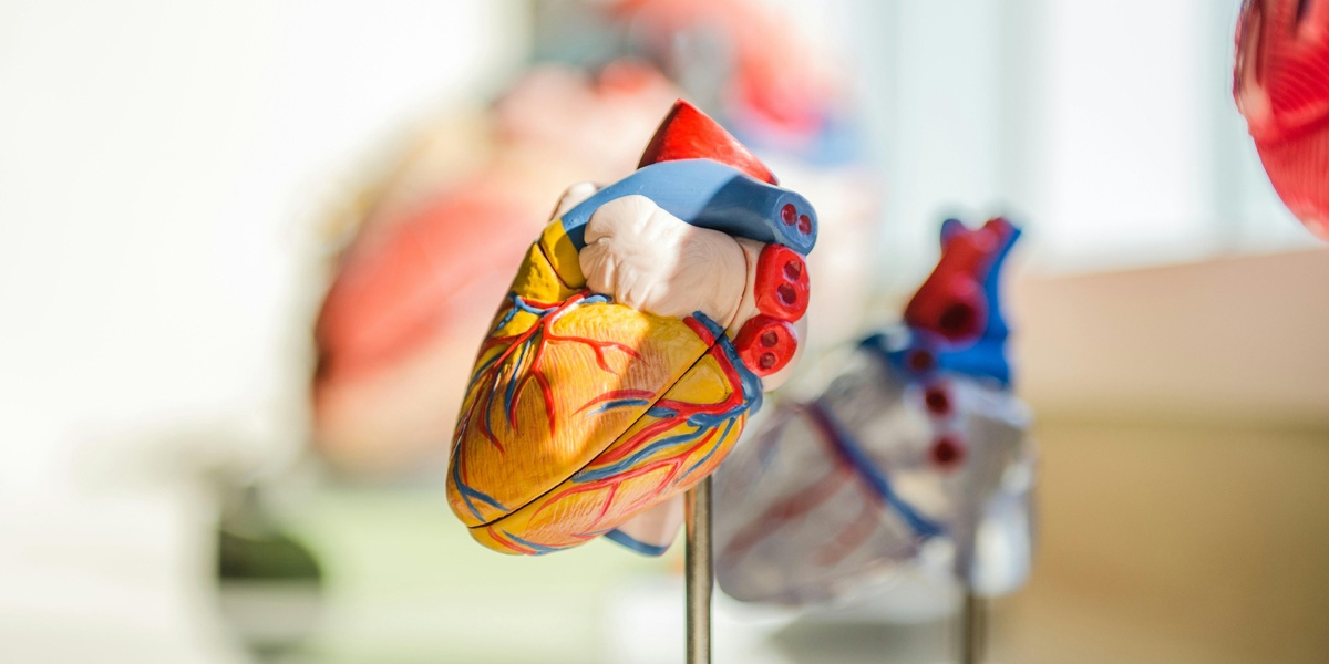
Left coronary artery (LCA)
Arises from aortic root and divides into LAD and circumflex; supplies majority of left ventricle. LCA disease causes large myocardial infarctions; critical in cardiac surgery and angioplasty.
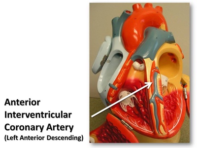
Left anterior descending artery (LAD)
Major LCA branch running in anterior interventricular sulcus supplying large anterior LV territory. “Widowmaker” lesions cause extensive myocardial infarction; primary PCI target in acute MI.
Left circumflex artery (LCx)
Runs in the atrioventricular groove supplying lateral and posterior LV. Variations affect myocardial perfusion; disease causes ischemia and is treated with CABG or stenting.
Right coronary artery (RCA)
Supplies right atrium, right ventricle and inferior LV; gives PDA in many people. RCA occlusion causes inferior MI and conduction disturbances due to AV nodal supply.
Posterior descending artery (PDA)
Runs in posterior interventricular sulcus supplying inferior interventricular septum. Origin variability (RCA or LCx) determines coronary dominance and affects infarct patterns.
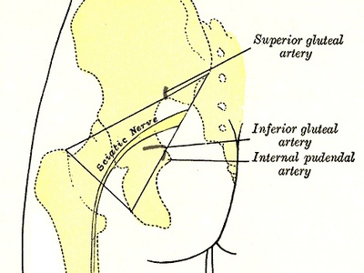
Superior gluteal artery
Largest branch of posterior division supplying gluteal muscles and hip capsule. Important in hip surgery and intramuscular injections; damage causes gluteal ischemia.

Inferior gluteal artery
Supplies gluteus maximus and posterior thigh; exits pelvis via greater sciatic foramen. Relevance in surgical approaches and trauma to pelvis and hip.
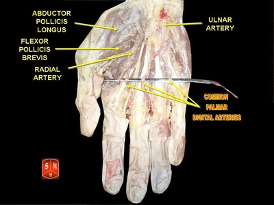
Common palmar digital arteries
Branches of the superficial palmar arch supplying adjacent sides of fingers. Important in hand trauma and digital ischemia; small caliber but clinically significant for perfusion.
Renal segmental arteries
Named intrarenal branches supplying discrete kidney segments; surgical importance in partial nephrectomy and embolization. Segmental infarcts follow occlusion due to end-artery nature.
Lumbar arteries
Paired segmental branches supplying posterior abdominal wall and spinal cord radicular branches. Important in abdominal surgery and trauma; may be a source of bleeding in retroperitoneal hemorrhage.
Median sacral artery
Small midline branch from aortic bifurcation supplying sacral region. Rarely clinically important but can be encountered in pelvic surgery or aortic aneurysm repair.
Superior rectal artery
Terminal branch of IMA supplying upper rectum and forming anastomoses with middle rectal arteries. Hemorrhoids and rectal bleeding can involve this vessel; ligated in some colorectal surgeries.
Inferior epigastric artery
Arises from external iliac supplying lower anterior abdominal wall and participating in collateral circulation. Important in hernia repairs and laparoscopic port placement to avoid bleeding.
Superior pancreaticoduodenal artery
Arises from gastroduodenal supplying pancreatic head and duodenum; anastomoses with inferior pancreaticoduodenal from SMA. Involved in pancreatic and duodenal bleeding management.
Right colic artery
Supplies ascending colon and hepatic flexure; important in colectomy planning. Variable origin makes surgical identification important to preserve colonic perfusion.
Middle colic artery
Supplies transverse colon via central mesocolon; crucial for maintaining colonic blood flow during colorectal resections. Anastomoses form marginal artery of Drummond.
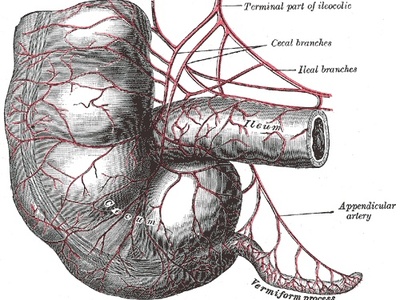
Ileocolic artery
Supplies terminal ileum, cecum and appendix; clinically significant in appendicitis surgery and right hemicolectomy because it supplies appendix base.
Left colic artery
Supplies descending colon and anastomoses with middle colic to maintain marginal artery. Important in colorectal resections and risk area for ischemic colitis.
Internal maxillary artery
Deep terminal branch supplying nasal cavity, palate, teeth and infra-temporal structures. Involved in severe epistaxis and facial trauma; embolization often targets branches here.
Posterior auricular artery
Small branch supplying posterior auricle and scalp; relevant in head and neck surgery and reconstructive flaps.

Superior laryngeal artery
Branch supplying laryngeal mucosa and muscles; important in thyroid and laryngeal surgery to preserve voice and prevent bleeding.
External pudendal artery
Supplies superficial perineal skin and external genitalia; encountered in hernia repairs and superficial perineal procedures.