Veins thread throughout the body, carrying deoxygenated blood back to the heart and serving as landmarks for imaging, lines and surgery. Knowing where a vein sits and how big it is helps clinicians choose access sites, interpret scans, and anticipate complications — from peripheral bruising to significant central venous issues.
There are 20 Examples of Veins, ranging from the Axillary vein to the Superior vena cava. For each entry I list the data in three columns: Location (region),Typical diameter (mm),Clinical note to make comparisons quick and practical — you’ll find below.
Which veins on the list are most often used for medical access and why?
Central access commonly uses the internal jugular, subclavian and femoral veins because of size, accessibility and predictable anatomy; peripheral lines typically use basilic or cephalic veins. The Superior vena cava is the main central conduit referenced after catheter placement, while the Axillary vein often serves as an alternative for peripheral or midline access.
How should vein diameter and location influence clinical decisions?
Vein diameter guides device choice (smaller veins limit catheter size) and risk assessment (larger central veins tolerate long-term lines better). Location affects infection and thrombosis risk, with central veins requiring stricter sterility and monitoring; use the columned data below to match clinical needs to anatomic characteristics.
Examples of Veins
| Name | Location (region) | Typical diameter (mm) | Clinical note |
|---|---|---|---|
| Superior vena cava | Thorax/upper chest | 20-28 | Central venous access; SVC syndrome possible |
| Inferior vena cava | Abdomen/retroperitoneum | 20-30 | Thrombosis and compression from masses; major central venous flow |
| Internal jugular vein | Neck | 10-18 | Common site for central lines and volume assessment |
| External jugular vein | Neck (superficial) | 4-8 | Visible with straining; used for peripheral access occasionally |
| Subclavian vein | Below clavicle | 8-12 | Route for central lines; risk of pneumothorax if misplaced |
| Axillary vein | Axilla/upper arm | 6-12 | Important in arm DVT and central access complications |
| Cephalic vein | Lateral forearm/arm (superficial) | 2-4 | Common for cannulation and pacemaker lead access |
| Basilic vein | Medial forearm/arm (superficial) | 3-6 | Used for phlebotomy and surgical access |
| Median cubital vein | Anterior elbow (antecubital) | 2-3 | Usual site for blood draws and IV insertion |
| Brachial vein | Arm (deep) | 4-6 | Deep arm vein; can thrombose or be catheterized |
| Azygos vein | Right posterior thorax | 6-8 | Important collateral with IVC obstruction; seen on imaging |
| Portal vein | Hepatoduodenal ligament/abdomen | 8-12 | Central in portal hypertension and variceal bleeding risk |
| Hepatic veins | Liver to IVC | 6-10 | Obstruction causes Budd–Chiari syndrome; affects liver outflow |
| Renal vein | Kidney to IVC | 4-8 | Left renal vein receives gonadal vein; relevant in surgery |
| Gonadal vein | Pelvis (testicular/ovarian) | 2-4 | Varicocele (left) and pelvic congestion; surgical ligation sometimes |
| Common iliac vein | Pelvis | 10-14 | Site for DVT and pelvic venous drainage |
| Femoral vein | Thigh (deep) | 8-12 | Major DVT site and used for vascular access |
| Great saphenous vein | Medial leg (superficial) | 3-5 | Often varicosed; harvested for bypass grafts |
| Popliteal vein | Behind knee | 6-8 | Important in DVT after knee injury or surgery |
| Superior sagittal sinus | Midline cranial vault | 6-12 | Sinus thrombosis causes headache, raised intracranial pressure |
Images and Descriptions
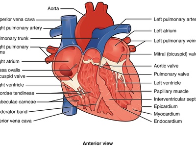
Superior vena cava
A large central vein returning blood from the head, neck, upper limbs, and chest to the right atrium; notable for central venous access, superior vena cava syndrome from obstruction, and as a landmark in many thoracic procedures.
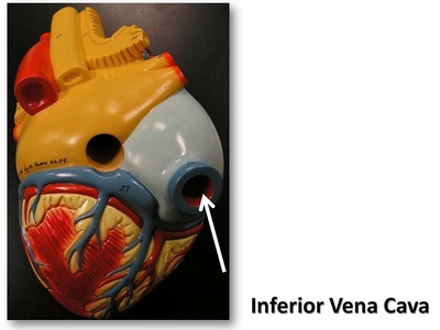
Inferior vena cava
The large abdominal vein returning blood from the pelvis, lower limbs, and abdominal organs to the right atrium; central in venous return and commonly involved in deep vein thrombosis, compression by tumors, and major vascular surgery.
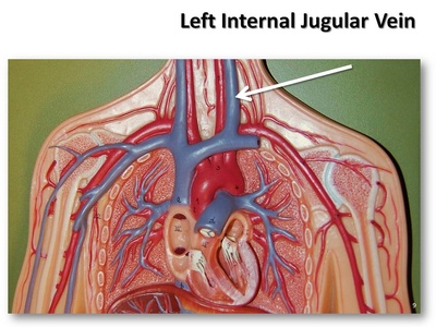
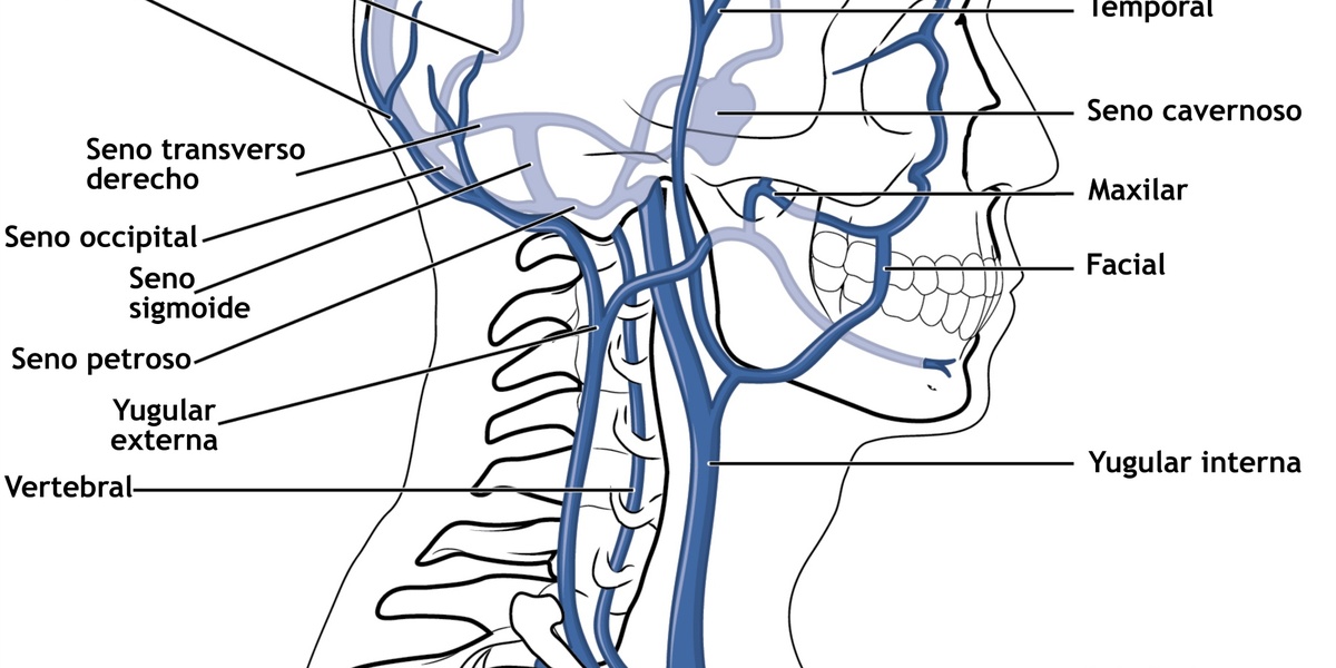
Internal jugular vein
Major neck vein that drains the brain, face, and neck into the brachiocephalic veins; commonly used for central lines, ultrasound assessment of volume status, and may thrombose in infections or after catheterization.
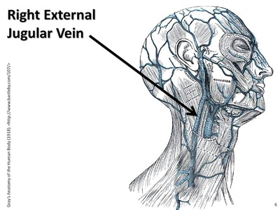
External jugular vein
Superficial neck vein draining exterior scalp and face into the subclavian vein; often visible when coughing or straining and used for peripheral venous access or assessment of venous pressure and occasionally cannulated in emergencies.
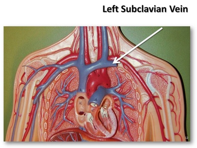
Subclavian vein
Large vein under the clavicle receiving blood from the arm via the axillary vein and draining into the brachiocephalic vein; central line route with risk of pneumothorax during access if misplaced.
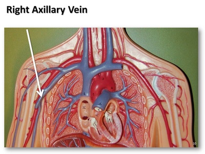
Axillary vein
Deep vein of the axilla formed by brachial veins and cephalic drainage; it continues as the subclavian vein and is important in central access complications, arm deep vein thrombosis, and regional infection spread.
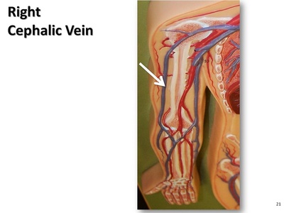
Cephalic vein
A superficial lateral arm vein running from the hand to the shoulder into the axillary/subclavian system; often used for cannulation, pacemaker lead access, and visible under the skin in many people.
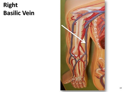
Basilic vein
Superficial medial arm vein that drains the hand and forearm into the brachial veins; commonly used for phlebotomy and forms a deep connection for arm venous return and surgical access.
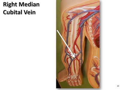
Median cubital vein
A superficial forearm/elbow vein connecting cephalic and basilic veins; the usual site for blood draws and peripheral IV insertion because of accessibility and clinicians choose it for ease and lower complication risk.
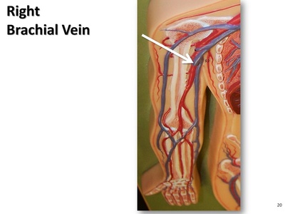
Brachial vein
Paired deep veins accompanying the brachial artery in the arm, draining forearm and hand blood toward the axillary vein; commonly compressed in catheterization and thrombosis of deep arm veins procedures.
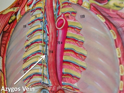
Azygos vein
Thoracic vein running along the right side of the vertebral column, draining posterior chest wall and mediastinal veins into the SVC; important collateral pathway with IVC obstruction and imaging assessment.
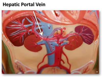
Portal vein
Large vessel carrying nutrient-rich blood from the gastrointestinal tract and spleen to the liver for processing; central in liver disease, portal hypertension, and variceal bleeding risk and crucial in surgical planning and imaging.
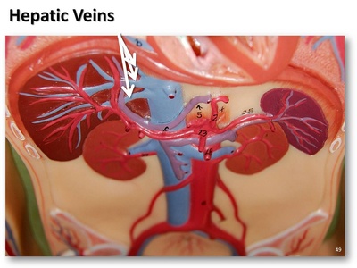
Hepatic veins
Veins that drain blood from the liver into the IVC, typically three main branches (right, middle, left); obstruction causes congestion, Budd–Chiari syndrome, and impaired liver outflow often requiring urgent care.

Renal vein
Paired veins draining each kidney into the IVC, carrying filtered blood; left renal vein is longer and receives the left gonadal vein, important in varicocele, renal surgery, and imaging.
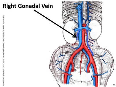
Gonadal vein
Testicular or ovarian vein draining gonads to the renal vein (left) or IVC (right); relevance in varicoceles in men and pelvic congestion in women and surgical ligation considerations during interventions.

Common iliac vein
Formed by internal and external iliac veins, the common iliac veins join to form the IVC; they drain the pelvis and lower limbs and are sites for deep vein thrombosis.
Femoral vein
Deep thigh vein continuing from the popliteal vein into the external iliac vein; major conduit for leg venous return and common site for DVT and vascular access and for endovascular procedures.
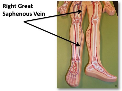
Great saphenous vein
Longest superficial vein running from the foot medial side to the femoral vein at the groin; commonly used for bypass grafts and frequently varicosed in many adults causing pain and leg swelling.

Popliteal vein
Deep vein behind the knee formed by tibial and fibular veins, continuing to the femoral vein; important in DVT diagnosis and at risk after knee injury or surgery or procedures.
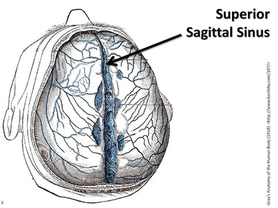
Superior sagittal sinus
A major midline dural venous sinus along the top of the brain draining cortical veins to the confluence of sinuses; relevant in sinus thrombosis and elevated intracranial pressure and head pain.