The forearm does the heavy lifting behind many everyday actions — from gripping a coffee cup to stabilizing the wrist during a push-up. Understanding its muscles makes it easier to diagnose pain, improve strength, or follow anatomy while studying or training.
There are 20 Muscles of the Forearm, ranging from Abductor pollicis longus to Supinator. For each muscle, the Compartment,Primary action,Innervation are listed in columns to make comparisons quick and clear — you’ll find below.
Which forearm muscles control wrist extension?
Wrist extension is mainly performed by the posterior compartment muscles, especially the extensor carpi radialis longus, extensor carpi radialis brevis, and extensor carpi ulnaris; these stabilize and lift the hand at the wrist and are typically innervated by branches of the radial nerve.
How can I reduce forearm pain and strengthen these muscles safely?
Begin with low-resistance, high-repetition exercises like wrist curls and finger extensions, add eccentric training for tendon health, and prioritize rest, gradual progression, and proper technique; if pain persists or is severe, get evaluated to rule out nerve entrapment or tendon injury.
Muscles of the Forearm
| Name | Compartment | Primary action | Innervation |
|---|---|---|---|
| Pronator teres | Anterior – superficial | Forearm pronation, assists elbow flexion | Median nerve (C6–7) |
| Flexor carpi radialis | Anterior – superficial | Wrist flexion, radial deviation | Median nerve (C7–8) |
| Palmaris longus | Anterior – superficial | Wrist flexion, tenses palmar aponeurosis | Median nerve (C7–8) |
| Flexor carpi ulnaris | Anterior – superficial | Wrist flexion, ulnar deviation | Ulnar nerve (C8–T1) |
| Flexor digitorum superficialis | Anterior – intermediate | PIP flexion of digits 2–5, wrist flexion | Median nerve (C7–T1) |
| Flexor digitorum profundus | Anterior – deep | DIP flexion of digits 2–5, grips | Lateral half: anterior interosseous (median); medial half: ulnar nerve |
| Flexor pollicis longus | Anterior – deep | Thumb IP flexion, grip support | Anterior interosseous nerve (median) |
| Pronator quadratus | Anterior – deep | Forearm pronation, DRUJ stabilization | Anterior interosseous nerve (median) |
| Brachioradialis | Posterior – superficial | Elbow flexion (best in mid-pronation) | Radial nerve (C5–7) |
| Extensor carpi radialis longus | Posterior – superficial | Wrist extension, radial deviation | Radial nerve (C6–7) |
| Extensor carpi radialis brevis | Posterior – superficial | Wrist extension, stabilizes wrist for finger extension | Deep branch of radial nerve (posterior interosseous) |
| Extensor digitorum | Posterior – superficial | Extension of digits 2–5 at MCP, assists wrist extension | Posterior interosseous nerve (radial) |
| Extensor digiti minimi | Posterior – superficial | Extension of little finger (digit 5) | Posterior interosseous nerve (radial) |
| Extensor carpi ulnaris | Posterior – superficial | Wrist extension, ulnar deviation | Posterior interosseous nerve (radial) |
| Anconeus | Posterior – superficial | Assists elbow extension, stabilizes elbow | Radial nerve (C7–8) |
| Supinator | Posterior – deep | Forearm supination | Deep branch of radial nerve (posterior interosseous) |
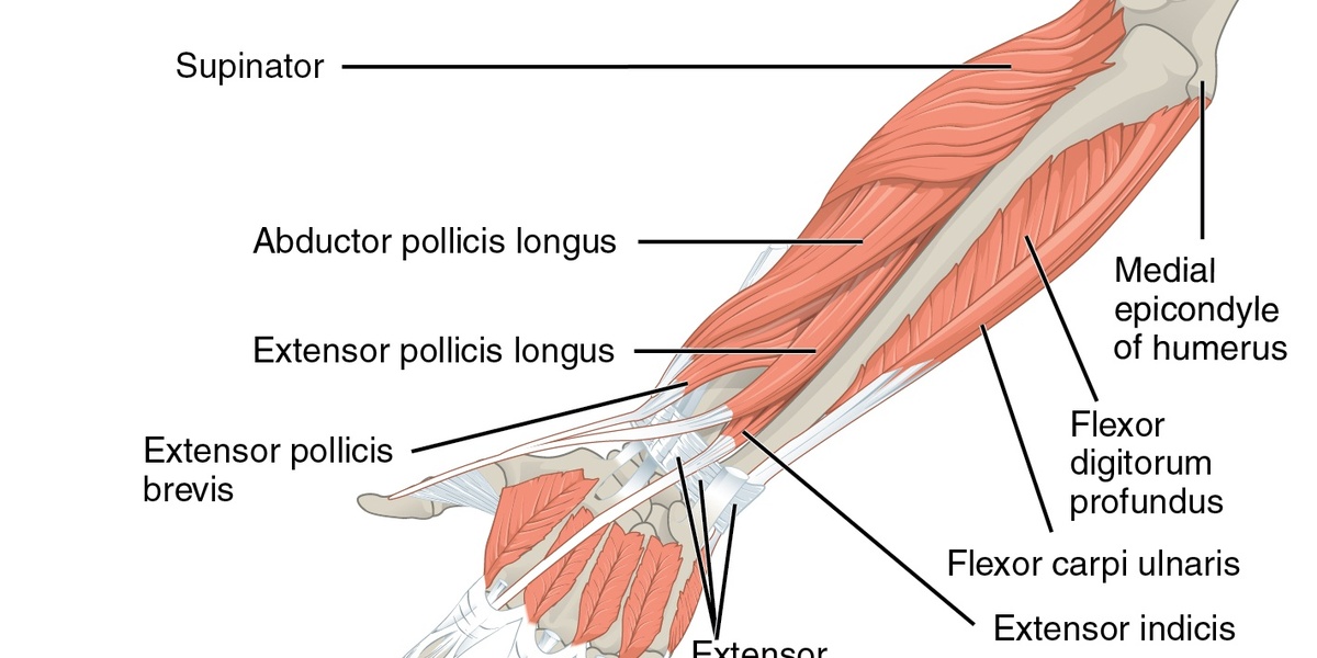
| Abductor pollicis longus | Posterior – deep | Thumb abduction, radial deviation of wrist | Posterior interosseous nerve (radial) |
| Extensor pollicis brevis | Posterior – deep | Thumb MCP extension, assists CMC abduction | Posterior interosseous nerve (radial) |
| Extensor pollicis longus | Posterior – deep | Thumb IP extension, assists wrist extension | Posterior interosseous nerve (radial) |
| Extensor indicis | Posterior – deep | Extension of index finger, independent pointing | Posterior interosseous nerve (radial) |
Images and Descriptions
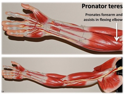
Pronator teres
Originates from medial epicondyle of humerus and coronoid process of ulna, inserts on lateral radius. Pronates the forearm and assists elbow flexion. Clinically relevant: pronator teres syndrome can compress the median nerve causing forearm pain and hand numbness.
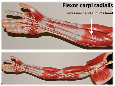
Flexor carpi radialis
Originates at medial epicondyle and inserts on base of 2nd metacarpal. Flexes and abducts the wrist, aiding grip. Clinically: overuse can cause wrist pain; tendon visible for pulse and sometimes used in grafts.
Palmaris longus
Originates from medial epicondyle and inserts into the palmar aponeurosis. Weak wrist flexor that tenses the palm; muscle belly is often small or absent. Clinically useful: tendon commonly harvested for grafts; absence is usually asymptomatic.
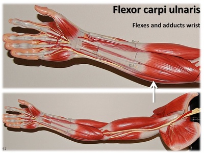
Flexor carpi ulnaris
Originates from medial epicondyle and olecranon, inserts on pisiform and 5th metacarpal. Powerful ulnar wrist flexor and adductor. Clinical note: close relation to the ulnar nerve; injury or entrapment can impair grip and ulnar deviation.

Flexor digitorum superficialis
Arises from humeroulnar head and radial head, inserting on middle phalanges of digits 2–5. Flexes PIP joints and assists wrist flexion. Clinically important in flexor tendon injuries and can be involved in tenosynovitis.

Flexor digitorum profundus
Originates on proximal ulna and interosseous membrane, inserting on distal phalanges 2–5. Flexes DIP joints and supports firm grip. Clinical note: dual innervation affects testing and is vulnerable in deep forearm lacerations.
Flexor pollicis longus
Originates from the anterior radius and interosseous membrane, inserting on the distal phalanx of the thumb. Flexes the thumb IP joint and aids pinch. Clinically: anterior interosseous syndrome impairs thumb flexion and key pinch strength.
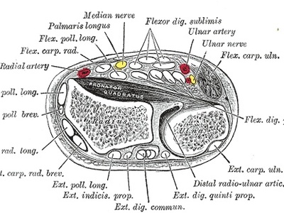
Pronator quadratus
A square muscle from distal ulna to distal radius that pronates the forearm and stabilizes the distal radioulnar joint. Clinically important: commonly preserved in proximal nerve injuries and relevant in distal radius fracture management.
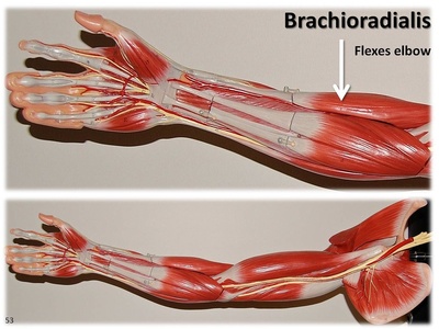
Brachioradialis
Originates on the lateral supracondylar ridge and inserts at the distal radius. Flexes the elbow especially with forearm in neutral. Clinical note: tendon is a landmark for radial nerve injury and often spared in posterior interosseous nerve palsy.
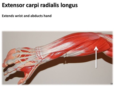
Extensor carpi radialis longus
Originates from lateral supracondylar ridge and inserts on base of 2nd metacarpal. Extends and abducts the wrist, stabilizing during hand use. Clinically implicated in lateral epicondylitis and overuse wrist pain.
Extensor carpi radialis brevis
Originates from the lateral epicondyle and inserts on base of 3rd metacarpal. Extends wrist and stabilizes it during finger extension. Clinically: major contributor to lateral epicondylitis (“tennis elbow”).
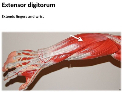
Extensor digitorum
Originates at the lateral epicondyle and inserts into extensor expansions of digits 2–5. Extends the MCP joints and helps extend PIP/DIP via the extensor hood. Clinically relevant in extensor tendon injuries and PIN palsy.
Extensor digiti minimi
Originates at the lateral epicondyle and inserts into the extensor expansion of the little finger. Specializes in extending digit 5 independently. Clinical note: can be injured or inflamed causing ulnar-sided dorsal wrist pain.
Extensor carpi ulnaris
Originates from lateral epicondyle and posterior ulna, inserting on base of 5th metacarpal. Extends and adducts the wrist. Clinically: tendonitis or subluxation causes ulnar-sided wrist pain and weakness with ulnar deviation.
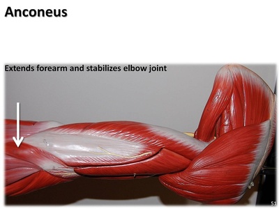
Anconeus
Small triangular muscle from lateral epicondyle to olecranon and proximal ulna. Assists triceps in elbow extension and stabilizes the elbow during rotation. Clinically minor but may be implicated in lateral elbow pain after trauma.
Supinator
Originates from lateral epicondyle and proximal ulna, inserting on lateral proximal radius. Powerful supinator of the forearm, especially with elbow extended. Clinical note: supinator (arcade of Frohse) can compress the deep radial nerve causing weakness.
Abductor pollicis longus
Arises from posterior radius, ulna and interosseous membrane; inserts on base of 1st metacarpal. Abducts thumb at CMC and assists wrist abduction. Clinically involved in De Quervain tenosynovitis causing radial wrist pain.
Extensor pollicis brevis
Originates on radius and interosseous membrane, inserting on proximal phalanx of thumb. Extends the thumb MCP and aids radial deviation. Clinically: tendon involved in De Quervain tenosynovitis producing radial wrist tenderness.
Extensor pollicis longus
Originates on the ulna and interosseous membrane, inserting on distal phalanx of thumb. Extends thumb at IP and MCP joints and helps wrist extension. Clinically: EPL tendon rupture may follow distal radius fractures.

Extensor indicis
Originates on distal ulna and interosseous membrane, inserting into the extensor expansion of the index finger. Permits independent extension of the index finger. Clinically useful as a tendon donor and affected in PIN lesions.