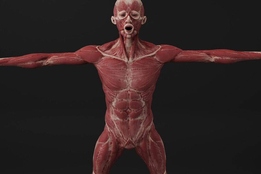
Muscle tissue powers everything from a quick reach to keeping blood flowing; you encounter it in limbs, the heart and inside organs. Knowing examples helps connect anatomy to real-world function, whether you’re studying physiology or planning training and rehab.
There are 20 Examples of Muscle Tissue, ranging from Biceps brachii to Vascular smooth muscle to illustrate the span of skeletal, cardiac and smooth types. For each entry you’ll find below the columns Tissue type, Location, Primary function so you can compare where each muscle sits and what it does—you’ll find below.
How do skeletal, cardiac and smooth muscles differ in structure and role?
Skeletal muscle (like the Biceps brachii) is striated and voluntary for movement and posture; cardiac muscle is striated with specialized junctions for rhythmic heart contractions; smooth muscle (for example, vascular smooth muscle) is non‑striated and involuntary, controlling vessel diameter and organ motility—structure ties directly to each muscle’s primary function.
Which examples should I focus on for exercise, injury risk, or health?
Focus on large skeletal muscles (quads, hamstrings, biceps) for exercise and injury prevention, and on smooth and cardiac examples when considering cardiovascular health or autonomic function; the table below helps prioritize by showing Tissue type, Location and Primary function for each muscle.
Examples of Muscle Tissue
| Name | Tissue type | Location | Primary function |
|---|---|---|---|
| Biceps brachii | Skeletal | Front of upper arm | Flexes elbow and supinates forearm |
| Triceps brachii | Skeletal | Back of upper arm | Extends the elbow for pushing actions |
| Deltoid | Skeletal | Shoulder | Abducts and rotates the arm |
| Pectoralis major | Skeletal | Chest (anterior thorax) | Adducts and medially rotates the arm |
| Rectus abdominis | Skeletal | Front of abdomen | Flexes the spine and stabilizes trunk |
| Quadriceps | Skeletal | Front of thigh | Extends the knee for standing and walking |
| Hamstrings | Skeletal | Back of thigh | Flexes knee and extends the hip |
| Gluteus maximus | Skeletal | Buttock/hip region | Extends and externally rotates the hip |
| Gastrocnemius | Skeletal | Calf (back of lower leg) | Plantarflexes ankle and aids propulsion |
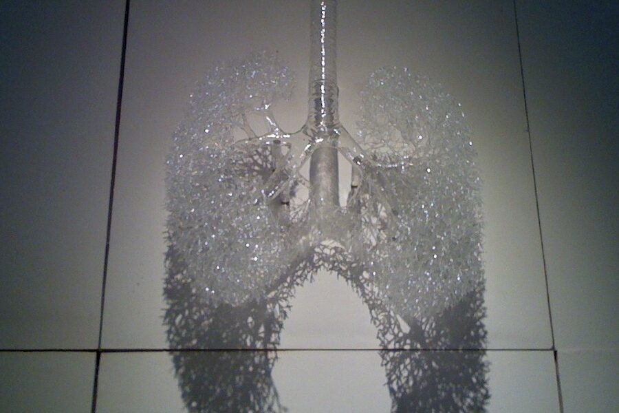
| Diaphragm | Skeletal | Between chest and abdomen | Primary muscle of inspiration (breathing) |
| Trapezius | Skeletal | Upper back and neck | Moves and stabilizes the shoulder blades |
| Latissimus dorsi | Skeletal | Mid to lower back | Adducts, extends, and internally rotates arm |
| Masseter | Skeletal | Side of jaw (angle of mandible) | Closes jaw for chewing |
| Sternocleidomastoid | Skeletal | Side of neck | Rotates and flexes the head and neck |
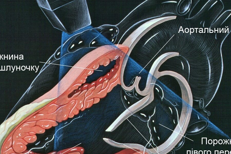
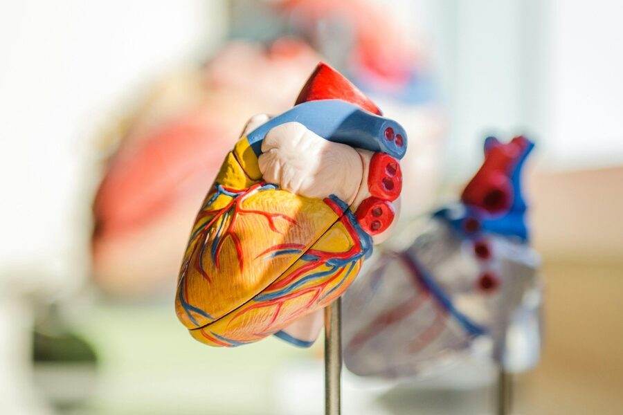
| Myocardium | Cardiac | Heart wall (ventricles) | Contracts to pump blood throughout body |
| Vascular smooth muscle | Smooth | Walls of arteries and arterioles | Regulates vessel diameter and blood pressure |
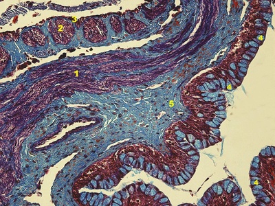
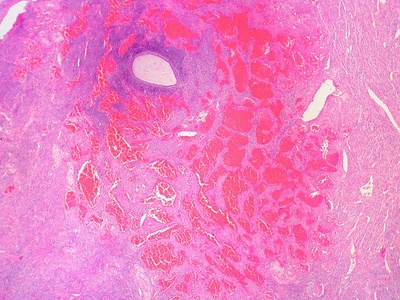
| Intestinal smooth muscle | Smooth | Walls of small and large intestine | Mixes and propels contents by peristalsis |
| Myometrium | Smooth | Wall of the uterus | Contracts during labor and menstruation |
| Detrusor muscle | Smooth | Urinary bladder wall | Contracts to expel urine and allow filling |
| Iris sphincter | Smooth | Iris of the eye | Constricts the pupil in bright light |
Images and Descriptions
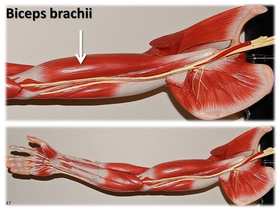
Biceps brachii
A prominent two-headed muscle on the front of the upper arm that flexes the elbow and assists forearm supination. Commonly visible during lifting and daily activities, it’s often used as a symbol of arm strength and is important for pulling motions.
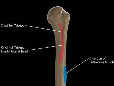
Triceps brachii
A large three-headed muscle on the back of the upper arm that straightens the elbow joint, enabling pushing actions. It balances the biceps during arm movement and plays a key role in activities like pushing, throwing, and rising from a seated position.
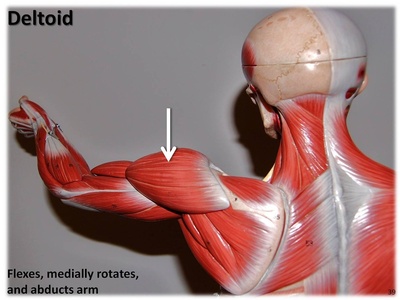
Deltoid
A triangular shoulder muscle that covers the joint and lifts the arm away from the body. Its three parts let you raise, rotate, and stabilize the arm in many directions, making it essential for reaching, throwing, and everyday overhead tasks.
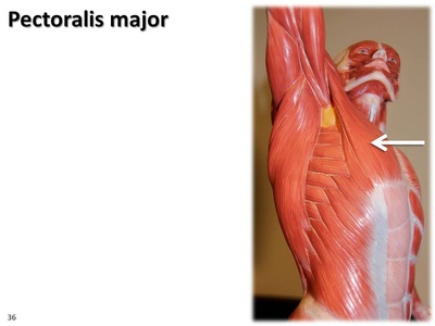
Pectoralis major
A broad chest muscle that brings the arm across the body and rotates it inward, contributing to pushing movements. Heavily used in activities like pushing doors, bench pressing, and hugging, it helps shape the front of the upper torso.
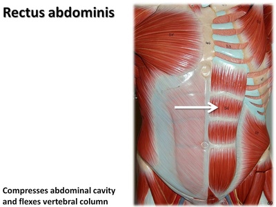
Rectus abdominis
The paired “six-pack” muscles running vertically on the front of the abdomen that flex the spine and stabilize the trunk. Important for posture, coughing, and core strength, they also protect internal organs and help with movements like sit-ups.

Quadriceps
A powerful group of four muscles on the front of the thigh that straightens the knee and supports standing and walking. Essential for running, jumping, and climbing, the quadriceps also stabilize the knee joint during weight-bearing activities.
Hamstrings
A group of muscles at the back of the thigh, including the biceps femoris, that bend the knee and extend the hip. Crucial for sprinting, walking, and posture, hamstrings help control leg swing and absorb impact during running.
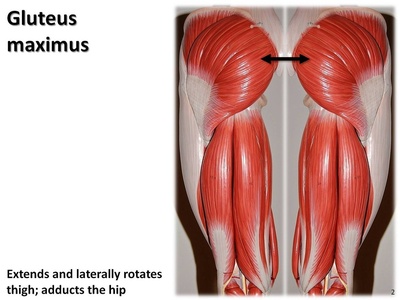
Gluteus maximus
The largest superficial muscle of the buttock that extends and externally rotates the hip, providing power for climbing, standing up, and running. It contributes significantly to posture and is a major source of strength and stability in the pelvis.
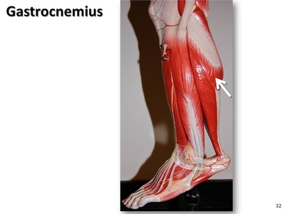
Gastrocnemius
The prominent calf muscle that, together with the soleus, points the toes (plantarflexes the ankle) and propels the body forward during walking and running. It’s visible behind the lower leg and contributes to balance and explosive movements.
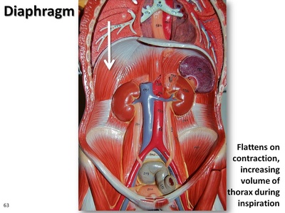
Diaphragm
The dome-shaped respiratory muscle separating chest and abdomen that contracts to expand the lungs and draw air in. As the primary muscle of breathing, it also assists with coughing, vomiting, and increasing abdominal pressure during lifting or childbirth.
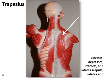
Trapezius
A large upper-back and neck muscle that moves and stabilizes the shoulder blades, helps tilt and turn the head, and supports arm movements. It plays a major role in posture, shrugging motions, and transferring forces from the arm to the trunk.
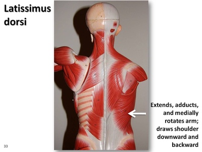
Latissimus dorsi
A broad, flat back muscle that pulls the arm down and backward and rotates it inward. Important for swimming, rowing, and climbing, it’s a powerful contributor to shoulder adduction and helps stabilize the lower back during lifting.
Masseter
A strong jaw muscle that closes the mouth and powers chewing, one of the most forceful muscles relative to its size. Located at the angle of the jaw, it’s essential for biting, grinding food, and contributing to facial shape and expression.
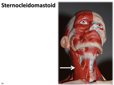
Sternocleidomastoid
A prominent neck muscle that tilts and rotates the head, and flexes the neck forward when both sides act together. It becomes noticeable with head movement, helps stabilize the head, and is often tense with stress or poor posture.
Myocardium
The thick muscular layer of the heart wall composed of cardiac muscle cells that contract rhythmically to pump blood. Highly resistant to fatigue and coordinated by electrical signals, the myocardium powers circulation to all organs and adapts to changing demands.
Vascular smooth muscle
Smooth muscle in blood vessel walls that contracts or relaxes to change vessel diameter and regulate blood pressure and flow. It responds to nervous signals, hormones, and local chemicals, playing a central role in circulation and temperature regulation.
Intestinal smooth muscle
Two layers of smooth muscle in the intestinal wall that mix and propel food by coordinated contractions called peristalsis. These muscles enable digestion, nutrient absorption, and movement of contents along the gastrointestinal tract.
Myometrium
The thick smooth-muscle layer of the uterus that contracts during labor to expel a baby and also during menstruation. Its powerful rhythmic contractions are under hormonal control and can change size dramatically during pregnancy.
Detrusor muscle
The smooth muscle forming the bladder wall that contracts to expel urine and relaxes to allow filling. Coordinated by the nervous system, the detrusor helps maintain continence and permits voluntary voiding when appropriate.
Iris sphincter
A circular smooth muscle in the iris that constricts the pupil in bright light or during near focus. Working with the dilator muscle, it helps control the amount of light entering the eye and contributes to depth of focus.