In 1839, the combination of improved microscopes and staining techniques launched modern histology—turning tissue slices into windows on disease. That leap from crude lenses to stained preparations made it possible for physicians to read cellular change and for surgeons to act on clearer evidence. Today those choices—what to look for, and how quickly to report—depend as much on instruments as on expertise.
From the familiar light microscope to modern molecular sequencers, a compact set of specialized devices underpins every diagnostic decision, affecting accuracy, turnaround time, and ultimately patient outcomes. Faster, standardized instruments reduce reporting variability; high-resolution imaging or molecular platforms can reveal targets for therapy that were invisible a decade ago. Below are eight essential instruments used in pathology and why each matters for diagnostic confidence, speed, or new diagnostic capabilities. Today, those early advances have become specialized instruments that labs rely on every day.
Microscopy and Imaging Tools
Microscopy remains the backbone of morphological diagnosis. Over the last century glass lenses gave way to automated optics, digital whole-slide scanners and, where needed, electron microscopes that reveal ultrastructure. These tools drive routine case sign-out, enable telepathology consults and feed AI algorithms that assist triage and quantitation.
Manufacturers such as Olympus and Leica supply clinical brightfield microscopes for day-to-day review, while Aperio/Leica, Hamamatsu and Philips produce whole-slide scanners used for digital archiving and remote review. Transmission electron microscopy (TEM), first demonstrated by Knoll and Ruska in 1931, remains indispensable for select diagnoses that require nanometer resolution.
1. Light Microscope (Brightfield)
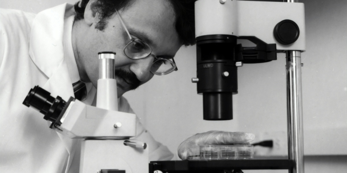
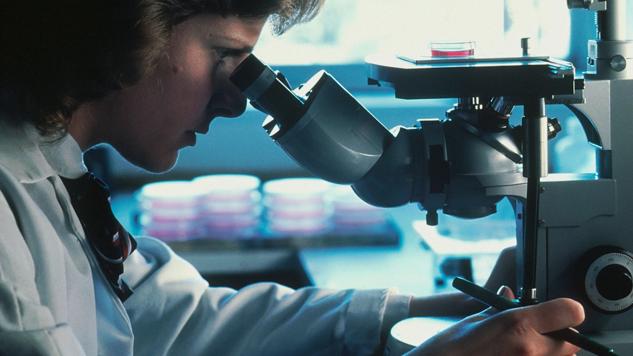
The brightfield light microscope is the core tool for routine histology: pathologists typically scan slides at low power, then examine critical fields at 40x–1000x (oil immersion) to assess cellular detail. Most surgical pathology workflows start at a histology microscope reviewing H&E stains.
Common clinical models include the Olympus BX53 and Leica DM2000. In many hospitals, a single pathologist will review 1–3 slides per case on average, while high-volume centers examine hundreds of slides each day, so ergonomics and reliable optics matter.
2. Electron Microscope (TEM/SEM)
Electron microscopy provides nanometer-scale resolution that light microscopy cannot match; TEM resolves structures from below 1 nm up to a few nanometers, revealing ultrastructural detail critical in niche diagnoses. Knoll and Ruska’s 1931 work established TEM as a tool for visualizing organelles and particles.
Clinically, EM is used in renal pathology to evaluate podocyte foot processes (for minimal change disease), in muscle biopsies and to identify viral particles when morphology is diagnostic. Limitations include high cost, specialized technicians and slower throughput—EM cases often take hours to days depending on lab capacity.
3. Whole-Slide Scanner / Digital Imaging
Whole-slide scanners convert glass slides into high-resolution digital images for viewing, sharing and algorithmic analysis. The FDA cleared specific WSI systems for primary diagnosis around 2017, which accelerated clinical adoption for telepathology and remote consults.
Popular platforms include the Leica Aperio AT2, Hamamatsu NanoZoomer and Philips IntelliSite. A single scanned slide can be several gigabytes, so storage and network planning become critical for labs adopting digital workflows. Digital slides also enable AI-assisted quantitation and teaching libraries.
Tissue Processing and Sectioning
Reliable diagnosis begins long before a slide reaches a microscope: tissue must be fixed, processed, embedded in paraffin or frozen, and sliced into thin sections. Standard paraffin sections are typically 3–5 µm thick; consistent thickness and processing remove many artifacts that would otherwise confound interpretation.
High-throughput labs use automated processors and embedding stations to handle dozens to 100+ cassettes per run, improving consistency and reducing technician-dependent variability. For rapid intraoperative decisions, cryosectioning provides speed at the cost of some morphological detail.
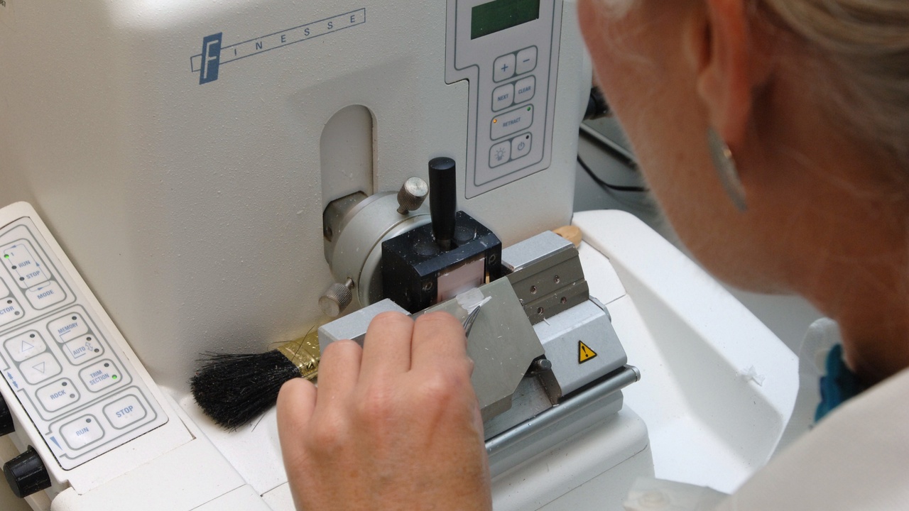
4. Microtome (Rotary / Automated)
A microtome slices paraffin-embedded tissue into sections typically 3–5 µm thick. Section thickness affects staining intensity and the ability to resolve nuclei and other structures, so precision matters for accurate grading and margin assessment.
Clinical units include the Leica RM2235 rotary microtome; automated modes and safety features (blade guards, programmable sectioning) help labs produce hundreds of sections per day in high-volume settings, while smaller labs may still rely on manual rotary instruments.
5. Cryostat (Frozen Section Unit)
Cryostats enable rapid frozen sections for intraoperative consultations, frequently delivering results within 10–20 minutes. They operate around −20°C to freeze tissue quickly for sectioning, which is essential for margin assessment in procedures like Mohs surgery or breast lumpectomy.
The Thermo Fisher CryoStar NX70 and Leica cryostats are common in clinical practice. The trade-off is lower morphological detail compared with paraffin sections and special considerations for safety and cryogen handling during use.
6. Tissue Processor and Paraffin Embedding Station
Automated tissue processors perform fixation, dehydration, clearing and paraffin infiltration in controlled cycles. Automation reduces variability between runs and increases throughput—some processors handle dozens to over 100 cassettes per program, with runs lasting several hours.
Platforms such as Leica Tissue-Tek VIP and Thermo Fisher Excelsior ES are standard in many labs. Consistent processing yields fewer artifacts, improves staining reproducibility and ensures reliable sections for downstream microtomy and IHC.
Staining, Safety and Molecular Diagnostic Instruments
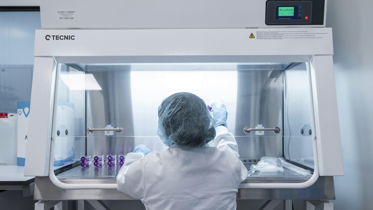
Beyond morphology, instruments used in pathology now include platforms for standardized staining, biosafety and molecular testing that expand diagnostic scope. Immunohistochemistry and molecular assays (PCR/NGS) provide predictive and prognostic information that directly guides therapy, while biosafety equipment protects staff and specimen fidelity.
Automation across staining and molecular workflows improves reproducibility and helps meet accreditation standards, enabling many labs to scale complex testing without sacrificing quality.
7. Automated Stainers and Immunohistochemistry (IHC) Platforms
Automated stainers standardize H&E and IHC protocols, cutting technician variability and manual labor. Systems such as the Dako Autostainer Link 48 and Roche’s Ventana Benchmark ULTRA can process dozens to hundreds of slides per run, depending on configuration.
These platforms support critical predictive tests—PD-L1 for immunotherapy selection and HER2 testing in breast cancer among them—and their consistency is central to reproducible scoring and compliance with lab accreditation requirements.
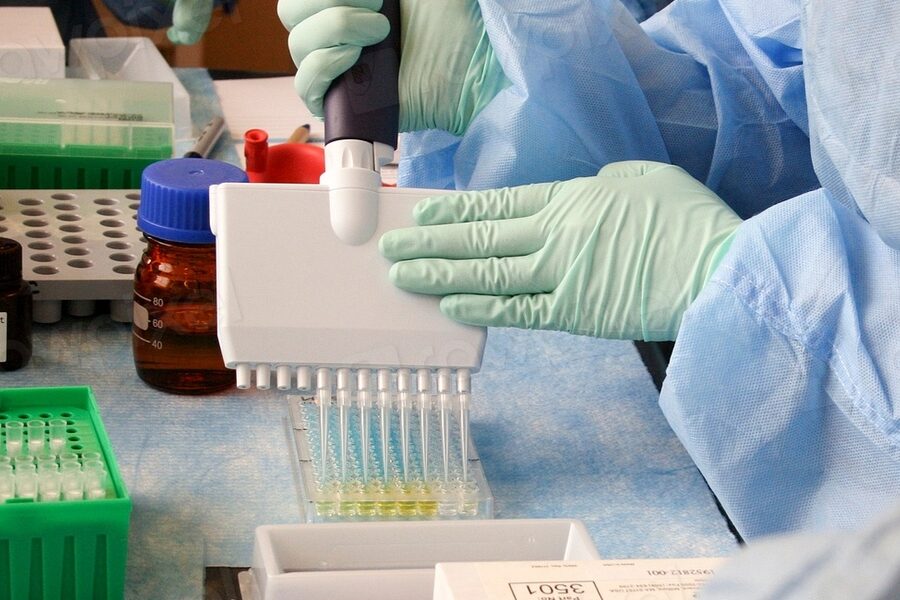
8. Molecular Diagnostics Platforms (PCR & NGS)
PCR and next-generation sequencing instruments identify genetic alterations that guide targeted therapy and infectious disease management. qPCR instruments like Thermo Fisher’s QuantStudio can return results in hours, while targeted NGS platforms such as Illumina MiSeq or NextSeq typically report within 1–7 days depending on panel size and batching.
Typical targeted NGS panels range from ~20 to 500 genes and generate millions of reads per run; a focused 50–100 gene panel is often used clinically to balance turnaround and depth. Platforms like Ion Torrent and Illumina have transformed oncology diagnostics by revealing actionable EGFR, BRAF and KRAS mutations and by supporting antimicrobial resistance testing.
Summary
- Light microscopy remains the foundational step in diagnosis—fast, inexpensive and still the first tool pathologists reach for.
- Digital whole-slide imaging (FDA milestones circa 2017) and automation enable remote review, AI assistance and centralized workflows—but require significant storage and IT planning.
- Accurate sectioning and processing (3–5 µm paraffin sections, rapid cryosections in 10–20 minutes) are essential upstream steps that determine slide quality.
- IHC stainers and molecular platforms (qPCR in hours; targeted NGS panels in ~2–7 days) provide predictive and actionable information that directly affects treatment choices.
- When evaluating lab investments or patient-care workflows, prioritize tools that improve reproducibility and shorten critical turnarounds while keeping specimen and staff safety front of mind.