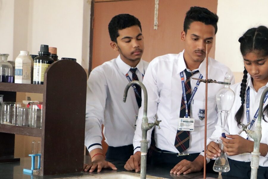
In 1884 Hans Christian Gram developed a staining technique that let scientists separate bacteria by cell wall type — a simple trick that transformed diagnosis and research. Practical, reliable laboratory instruments have done the same ever since, turning tiny, invisible organisms into clear signals clinicians and researchers can act on.
When microscopy and cultures are available, infectious disease diagnostics can cut diagnostic delay and improve outcomes; for example, rapid Gram-stain reads (results in about 30–60 minutes) often guide early treatment in hospitals. Tools matter for public health, medicine, food safety and basic research because they turn observations into data, and data into decisions.
This piece explains eight essential tools used in microbiology across three broad categories — microscopy and imaging; culturing and isolation; and molecular and analytical methods — showing what each does, why it matters, and real-world examples from clinics and labs.
Microscopy and Imaging Tools
1. Light Microscopy: Observing cell shape and stains
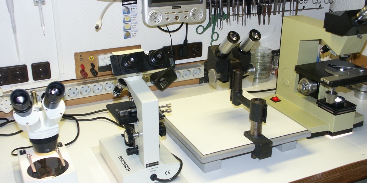
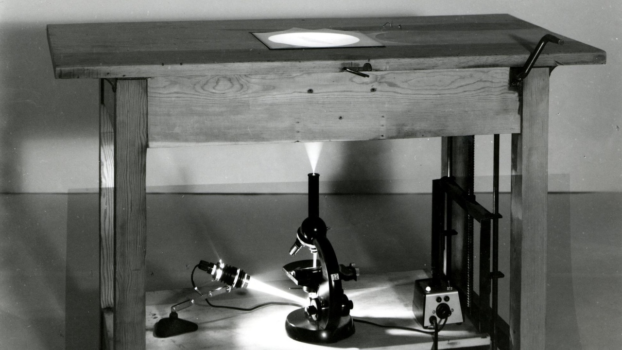
Light microscopes are the most common instruments in labs for viewing cells, Gram stains and simple wet mounts. Typical optical resolution sits around ~200 nm, which is plenty to assess cell shape, arrangement and staining patterns but falls short of seeing viral particles. Common magnifications used in clinical and teaching settings include 100x, 400x and 1,000x (the latter achieved with a 100x oil-immersion objective plus a 10x eyepiece).
Because Gram’s 1884 stain is still a go-to rapid test, many hospitals rely on compound microscopes from makers like Zeiss, Olympus or Leica for routine reads. Practical examples include malaria smears, quick Gram-stain reads in emergency rooms, and classroom labs where inexpensive Axioscope-style instruments teach fundamentals.
2. Fluorescence Microscopy: Specific labeling and diagnostics
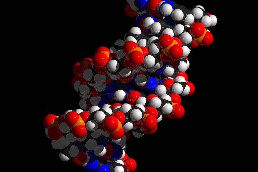
Fluorescence microscopy uses dyes or fluorescent proteins to label specific structures, raising specificity beyond what brightfield alone can provide. Common labels include GFP in research strains, DAPI for DNA, and Alexa dyes for antibody-based staining. Confocal systems (Leica, Nikon) add optical sectioning for clearer subcellular images.
Clinically, fluorescence enables immunofluorescence tests and FISH (fluorescent in situ hybridization) to detect particular pathogens or genes in patient and environmental samples. Many labs adopted fluorescence tools widely in the late 20th century, and some tuberculosis programs use fluorescent smear microscopy to reduce reading time per slide.
3. Electron Microscopy: Ultra-high resolution views
When nanometer-scale detail is required, electron microscopes come into play. Transmission EM (TEM) can resolve structures well below 1 nm, revealing internal ultrastructure, while scanning EM (SEM) provides high-resolution surface topology. That resolution is why JEOL and Thermo Fisher TEMs sit in major research centers.
EM is essential for viral morphology studies (think influenza or SARS‑CoV‑2 imaging) and materials–biology interfaces. The trade-offs are real: EM needs fixation, dehydration and heavy-metal staining, and prep can take hours to days, so it’s used when light microscopy lacks sufficient detail.
Culturing and Isolation Tools
4. Petri Dishes & Agar: Isolating colonies and counting CFUs
Petri dishes filled with agar let microbiologists isolate single colonies and perform standard plate counts to estimate colony-forming units (CFU/ml). A 90 mm plate with selective or differential media — MacConkey for Gram‑negative enterics, blood agar for hemolysis patterns — makes it straightforward to pick colonies for ID.
Plate counts are a workhorse in food safety and clinical labs: you plate, incubate (many bacteria show colonies in 24–48 hours), and count. Isolation into single colonies is how labs confirm pathogens and perform susceptibility testing, usually under BSL‑2 practices for common clinical cultures.
5. Incubators & Shakers: Controlling growth conditions
Incubators maintain temperature, humidity and sometimes CO2 (5% for many mammalian cell cultures), while orbital shakers supply aeration for liquid cultures. Typical settings include 37°C for many human pathogens and ~30°C for environmental isolates.
Thermo Scientific and Eppendorf make common incubators and shakers used in labs. Practical examples: growing E. coli for plasmid preparation (E. coli doubling time is about ~20 minutes under ideal conditions), culturing yeast, or incubating clinical specimens prior to plating.
6. Autoclaves & Sterilization: Ensuring aseptic work
Autoclaves are the go-to for sterilizing media, glassware and instruments. Standard parameters are 121°C at 15 psi for 15–20 minutes; that reliably inactivates most microbes and is required for treating biohazardous waste in clinical and research settings.
Labs also use filtration for heat‑sensitive solutions, dry‑heat ovens for certain glassware, and sterile single‑use loops or plates to reduce cross‑contamination. Bench‑top autoclaves are common in teaching and hospital labs, and proper sterilization is central to protecting staff and preserving experimental validity.
Molecular and Analytical Tools
7. PCR & Thermal Cyclers: Detecting DNA and RNA quickly
Polymerase chain reaction (PCR), developed by Kary Mullis in 1983, amplifies specific DNA sequences and transformed diagnostic microbiology. Quantitative PCR (qPCR) adds measurement, letting labs quantify targets; typical runs take about 1–2 hours depending on protocol and instrument.
Among the most impactful tools used in microbiology are thermal cyclers from Applied Biosystems and Bio‑Rad paired with extraction kits (QIAGEN) that prepare nucleic acids. High-sensitivity assays can detect down to single‑digit copies in optimized workflows, which is why qPCR played a central role in SARS‑CoV‑2 testing during 2020.
8. Gel Electrophoresis & Spectrophotometry: Visualizing and measuring biomolecules
Gel electrophoresis separates DNA and RNA by size so you can check PCR product length and integrity. Spectrophotometers and fluorometers quantify nucleic acids and proteins: a Nanodrop gives quick A260/A280 purity ratios (≈1.8–2.0 for clean DNA), while a Qubit fluorometer measures low concentrations more accurately in ng/µL.
Typical lab setups include Bio‑Rad gel rigs and simple spectrophotometers for routine QC before sequencing or cloning. These instruments are small but critical: they help detect failed preps early, saving time and reagents downstream.
Summary
- Tools translate microscopic biology into actionable results for patient care, food safety and research — from Gram’s 1884 stain up to PCR (1983) and qPCR workflows.
- Both low‑tech items (Petri dishes, microscopes, autoclaves at 121°C/15 psi for 15–20 minutes) and high‑tech instruments (TEMs, thermal cyclers, fluorometers) are essential parts of a functioning lab.
- Good sterilization, controlled incubation (37°C for many human pathogens) and basic QC steps (A260/A280, gel checks) prevent errors and protect staff and patients.
- Investing in training, maintenance and reliable tools used in microbiology pays off: faster diagnostics, better surveillance and safer labs — so support lab infrastructure and hands‑on expertise.